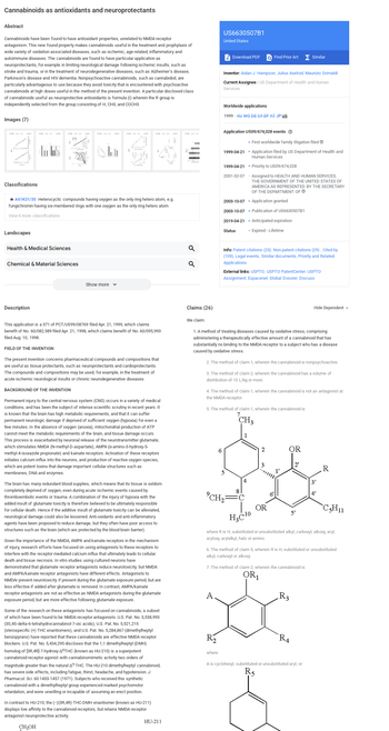
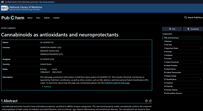
Napalm Death / Primitive Man / Ischemic / Sago / Pretty Mouth

Lee's Palace, Tuesday, June 2 at 07:00 PM EDT

https://toronto.askapunk.net/event/napalm-death-primitive-man-ischemic-sago-pretty-mouth

Blood Test Pinpoints Stoke Type In Ambulance: Hand-held device rapidly determines if a stroke is hemorrhagic or ischemic permitting earlier therapy. #stroke #gfap #hemorrhagic #ischemic #ambulance #prehospital
https://www.instagram.com/drhowardsmithreports/reel/DGWAQ9WPyMT/
Howard G. Smith MD, AM on Instagram: "Blood Test Pinpoints Stoke Type In Ambulance A handheld device carried in ambulances can rapidly determine whether a stroke is caused by a blocked brain blood vessel or by one that is bleeding. German neurologists have just reported this advance at this year’s International Stroke Conference. The device determines blood levels of glial fibrillary acidic protein or GFAP for short. Using it to test 353 suspected stroke victims, the researchers found that those with high GFAP levels were most likely to have hemorrhagic or bleeding strokes while those with low GFAP levels below 30 pg/ml had ischemic strokes due to clotted or blocked blood vessels. Knowing this information earlier permits a prompt initiation of appropriate therapy: clot dissolvers or clot removal for ischemic strokes or stringent blood pressure control for hemorrhagic strokes. This could drastically change pre-hospital care for strokes…….someday soon. https://newsroom.heart.org/news/blood-test-may-detect-stroke-type-before-hospital-arrival-allowing-faster-treatment #stroke #gfap #hemorrhagic #ischemic #ambulance #prehospital"

0 likes, 0 comments - drhowardsmithreports on February 21, 2025: "Blood Test Pinpoints Stoke Type In Ambulance A handheld device carried in ambulances can rapidly determine whether a stroke is caused by a blocked brain blood vessel or by one that is bleeding. German neurologists have just reported this advance at this year’s International Stroke Conference. The device determines blood levels of glial fibrillary acidic protein or GFAP for short. Using it to test 353 suspected stroke victims, the researchers found that those with high GFAP levels were most likely to have hemorrhagic or bleeding strokes while those with low GFAP levels below 30 pg/ml had ischemic strokes due to clotted or blocked blood vessels. Knowing this information earlier permits a prompt initiation of appropriate therapy: clot dissolvers or clot removal for ischemic strokes or stringent blood pressure control for hemorrhagic strokes. This could drastically change pre-hospital care for strokes…….someday soon. https://newsroom.heart.org/news/blood-test-may-detect-stroke-type-before-hospital-arrival-allowing-faster-treatment #stroke #gfap #hemorrhagic #ischemic #ambulance #prehospital".

Instagram

Iniciar tratamiento con #anticoagulante oral en los primeros 4 días después de un #EVC #isquémico en un paciente con #FibrilaciónAuricular es seguro, y beneficia más al paciente que iniciarlo después (5-14 días).

#OPTIMAS #CATALYST #Ischemic #Stroke #Anticoagulation #AtrialFibrillation

https://www.thelancet.com/journals/lancet/article/PIIS0140-6736(24)02197-4/fulltext

Looking for any other stroke or CVA survivors who run (or try to run). Hoping to start a community of us or join one already going #CVA #strokesurvivor #Ischemic #CVI #disabledrunner #running

Ischemic announce new album, title track streaming now

Ischemic, Ontario’s premier death/doom metal act, is thrilled to announce the release of their new single, the title track from their upcoming album, Condemned to the Breaking Wheel. This latest offering gives fans a first taste of what to expect from their highly anticipated full-length album due for release on April

https://www.moshville.co.uk/news/2024/02/ischemic-announce-new-album-title-track-streaming-now/

#News #Ischemic

Promising Findings – Scientists Successfully Revive Lost Brain Function Following Stroke

Researchers have successfully revived brain function in mice affected by stroke through the use of small molecules, which could potentially be developed into a stroke recovery therapy. “Communication between nerve cells in large parts of the brain changes after a stroke and we show that it can be

SciTechDaily
http://bit.ly/3THmLuZ
One month ago a group of whistleblowers sent the US NIH
a 113-pp. dossier about alleged fraud in a #Neurology lab at
Keck School of #Medicine of University of Southern #California Fraud was related to phase 2 of clinical trial of 3K3A-APC,a drug to prevent #Brain damage after #Ischemic stroke but that it could mean an advance in the treatment of #Alzheimer's disease too

Head-to-head comparison of #cardiac MR techniques pits parametric techniques against convential cardiac MR to visually differentiate #ischemic from non-ischemic #myocardial injury patterns. (Charlotte Jahnke et al.)

#EuropeanRadiology

🔗 https://link.springer.com/article/10.1007/s00330-023-09905-5

Differentiation of acute non-ST elevation myocardial infarction and acute infarct-like myocarditis by visual pattern analysis: a head-to-head comparison of different cardiac MR techniques - European Radiology

Objectives Parametric cardiac magnetic resonance (CMR) techniques have improved the diagnosis of pathologies. However, the primary tool for differentiating non-ST elevation myocardial infarction (NSTEMI) from myocarditis is still a visual assessment of conventional signal-intensity-based images. This study aimed at analyzing the ability of parametric compared to conventional techniques to visually differentiate ischemic from non-ischemic myocardial injury patterns. Methods Twenty NSTEMI patients, twenty infarct-like myocarditis patients, and twenty controls were examined using cine, T2-weighted CMR (T2w) and late gadolinium enhancement (LGE) imaging and T1/T2 mapping on a 1.5 T scanner. CMR images were presented in random order to two experienced fully blinded observers, who had to assign them to three categories by a visual analysis: NSTEMI, myocarditis, or healthy. Results The conventional approach (cine, T2w and LGE combined) had the best diagnostic accuracy with 92% (95%CI: 81–97) for NSTEMI and 86% (95%CI: 71–94) for myocarditis. The diagnostic accuracies using T1 maps were 88% (95%CI: 74–95) and 80% (95%CI: 62–91), 84% (95%CI: 67–93) and 74% (95%CI: 54–87) for LGE, and 83% (95%CI: 66–92) and 73% (95%CI: 53–87) for T2w. The accuracies for cine (72% (95%CI: 52–86) and 60% (95%CI: 38–78)) and T2 maps (62% (95%CI: 40–79) and 47% (95%CI: 28–68)) were significantly lower compared to the conventional approach (p < 0.001 and p < 0.0001). Conclusions The conventional approach provided a reliable visual discrimination between NSTEMI, myocarditis, and controls. The diagnostic accuracy of a visual pattern analysis of T1 maps was not significantly inferior, whereas the diagnostic accuracy of T2 maps was not sufficient in this context. Clinical relevance statement The ability of parametric compared to conventional CMR techniques to visually differentiate ischemic from non-ischemic myocardial injury patterns can avoid potentially unnecessary invasive coronary angiography and help to shorten CMR protocols and to reduce the need of gadolinium contrast agents. Key Points • A visual differentiation of ischemic from non-ischemic patterns of myocardial injury is reliably achieved by a combination of conventional CMR techniques (cine, T2-weighted and LGE imaging). • There is no significant difference in accuracies between visual pattern analysis on native T1 maps without providing quantitative values and a conventional combined approach for differentiating non-ST elevation myocardial infarction, infarct-like myocarditis, and controls. • T2 maps do not provide a sufficient diagnostic accuracy for visual pattern analysis for differentiating non-ST elevation myocardial infarction, infarct-like myocarditis, and controls.

SpringerLink