Lovely vacation with my folks on Mt Desert Island in Maine. Amazing spot on the water, I brought a little scope and had a blast looking at sea life. Even the lichen next to the house had tardigrades! #sciart #microscopy


Conference season is in full swing! 🎉
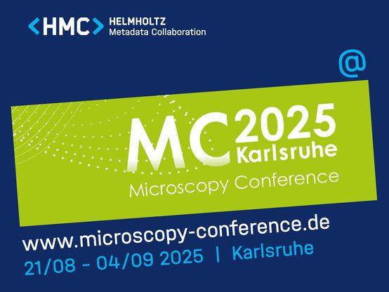
After #CoRDI2025 we’re off to the Microscopy Conference 2025 #MC2025 in Karlsruhe to talk about the #EMGlossary.
Join us on
🔹 Sunday, 31/08: for our presentation in Workshop 6 at 10:50h
and meet us
🔹 Wednesday, 03/09 at our poster (IM6.P10, 14:00–16:00) to learn more about the EM Glossary.
If you’ve seen our #WordoftheWeek series, come by & learn more about how to use or join the glossary. Let’s connect! 🤝
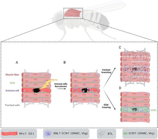
I have written another #preLight post, featuring the first work from the Boukhatmi lab.
Always fun to read a #fly paper, especially one with #microscopy too!
Check it out if you like injury repair and tissue/ECM remodelling.
Atomic Force Microscopy Nanoparticles for Precision Research | Molecular Imaging
#Atomic #Force #Microscopy nanoparticles offer unmatched accuracy in visualizing nanoscale structures. Molecular Imaging delivers powerful AFM tools that help researchers analyze particle size, morphology, and interactions. Our advanced imaging technology ensures reliable results for scientific innovation and cutting-edge discoveries in nanoscience.
Visit Now: https://miafm.com/nano-material/

Neurophotonics supports #OpenScience, transparency, #Reproducibility, and trust in research. Data sharing enables others to reuse your data for new discoveries increasing your impact!
https://www.spiedigitallibrary.org/journals/neurophotonics/author-guidelines#navBarAnchor
#neuroscience #neurophotonics #microscopy #fNIRS #OpticalImaging
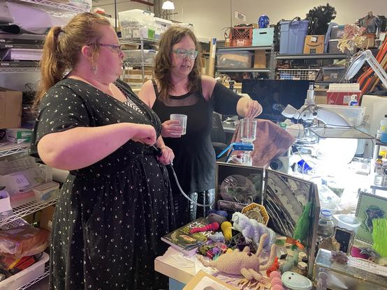
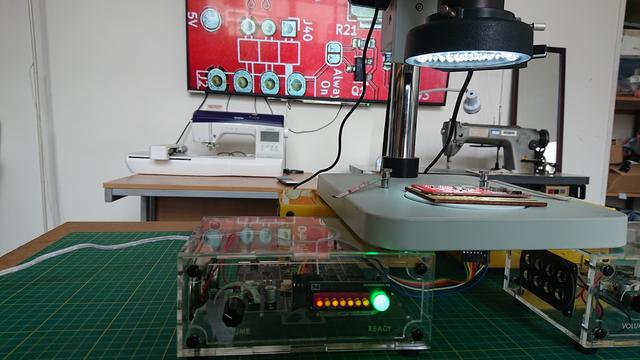
It worked quite nicely. Got some video and photos to share, so I'll write it up on the @museuminabox blog.
In the meantime, a sneak peek...
🔬📄 'Efficacy and Feasibility of Tissue-Clearing Technique and Three-Dimensional Imaging in the Human Gastrointestinal Tissues Using Illuminate Cleared Organs to Identify Target Molecules' - a Karger: #Gastroenterology article on #ScienceOpen -
🔗 https://www.scienceopen.com/document?vid=09d6ca4d-9131-4734-89df-86fae09bc8d7
#TissueClearing #3DImaging #LUCIDProtocol #Pathology #Microscopy
<p xmlns:xsi="http://www.w3.org/2001/XMLSchema-instance" dir="auto" id="d7337466e239"> <b> <i>Introduction:</i> </b> Tissue-clearing technology has shown potential for comprehensive structural and functional analysis through three-dimensional (3D) imaging of biological tissue. However, its effectiveness in human specimens remains insufficiently explored. In this study, we validated the illuminate cleared organs to identify target molecules (LUCID) protocol for human gastrointestinal specimens and demonstrated its utility in enhancing tissue transparency and 3D imaging. <b> <i>Methods:</i> </b> The gastrointestinal mucosa specimens resected via endoscopic submucosal dissection including the esophagus, stomach, duodenum, and colon were fluorescently stained and optically cleared using LUCID. Cleared specimens were imaged in 3D form by confocal laser scanning microscope, and the observable depth at any five points was measured and compared to non-cleared specimens, respectively. After clearing and imaging, the specimens were restored to the formalin-fixed paraffin-embedded form again, and conventional two-dimensional pathological evaluation using hematoxylin-eosin, Ki67, p53, and E-cadherin staining was performed to compare them with their preclearing state. <b> <i>Results:</i> </b> The observable depth was significantly extended after clearing for specimens from each organ (esophagus 228.3 ± 14.9 µm vs. 1,036.7 ± 62.9 µm, <i>p</i> < 0.05; stomach 115.2 ± 5.5 µm vs. 428.7 ± 15.9 µm, <i>p</i> < 0.05; duodenum 256.2 ± 9.5 µm vs. 787.0 ± 18.6 µm, <i>p</i> < 0.05, colon 113.9 ± 5.4 µm vs. 436.6 ± 18.5 µm, <i>p</i> < 0.05). The pathological evaluation after clearing revealed a preserved fine structure and staining and showed no apparent deformation, degeneration, or tissue damage compared with before clearing. <b> <i>Conclusions:</i> </b> The effectiveness of tissue clearing using LUCID on human gastrointestinal specimens was demonstrated, and the LUCID protocol had minimal impact on specimen morphology and staining. LUCID is expected to be a method that enables comprehensive structural analysis of human gastrointestinal mucosa and lesions that may avoid missing microscopic findings that may occur in split-face pathological assessment. </p>

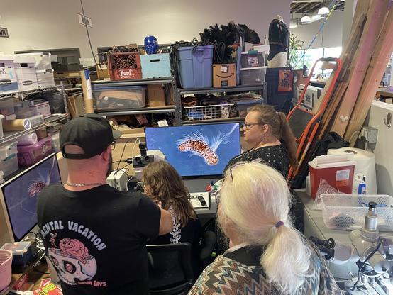
It worked quite nicely. Got some video and photos to share, so I'll write it up on the @museuminabox blog.
In the meantime, a sneak peek...