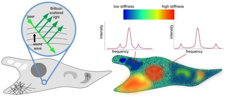
Neurophotonics supports #OpenScience, transparency, #Reproducibility, and trust in research. Data sharing enables others to reuse your data for new discoveries increasing your impact!
https://www.spiedigitallibrary.org/journals/neurophotonics/author-guidelines#navBarAnchor
#neuroscience #neurophotonics #microscopy #fNIRS #OpticalImaging
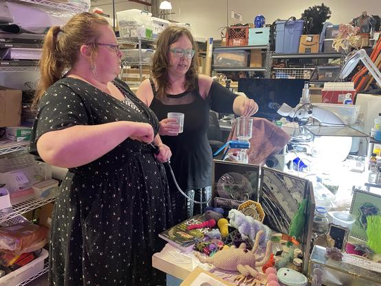
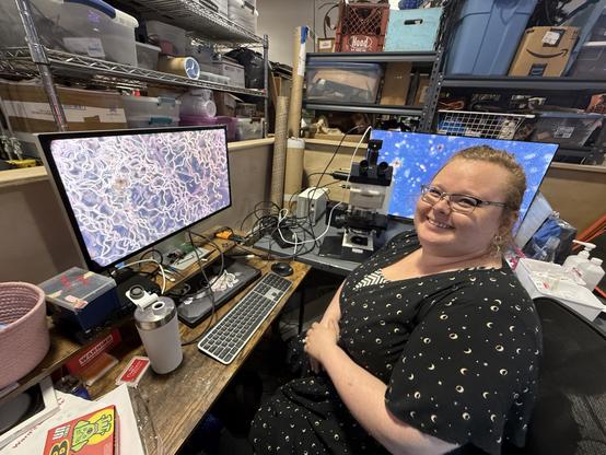
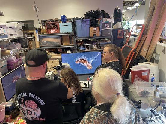
It worked quite nicely. Got some video and photos to share, so I'll write it up on the @museuminabox blog.
In the meantime, a sneak peek...
🔬📄 'Efficacy and Feasibility of Tissue-Clearing Technique and Three-Dimensional Imaging in the Human Gastrointestinal Tissues Using Illuminate Cleared Organs to Identify Target Molecules' - a Karger: #Gastroenterology article on #ScienceOpen -
🔗 https://www.scienceopen.com/document?vid=09d6ca4d-9131-4734-89df-86fae09bc8d7
#TissueClearing #3DImaging #LUCIDProtocol #Pathology #Microscopy
Efficacy and Feasibility of Tissue-Clearing Technique and Three-Dimensional Imaging in the Human Gastrointestinal Tissues Using Illuminate Cleared Organs to Identify Target Molecules
<p xmlns:xsi="http://www.w3.org/2001/XMLSchema-instance" dir="auto" id="d7337466e239"> <b> <i>Introduction:</i> </b> Tissue-clearing technology has shown potential for comprehensive structural and functional analysis through three-dimensional (3D) imaging of biological tissue. However, its effectiveness in human specimens remains insufficiently explored. In this study, we validated the illuminate cleared organs to identify target molecules (LUCID) protocol for human gastrointestinal specimens and demonstrated its utility in enhancing tissue transparency and 3D imaging. <b> <i>Methods:</i> </b> The gastrointestinal mucosa specimens resected via endoscopic submucosal dissection including the esophagus, stomach, duodenum, and colon were fluorescently stained and optically cleared using LUCID. Cleared specimens were imaged in 3D form by confocal laser scanning microscope, and the observable depth at any five points was measured and compared to non-cleared specimens, respectively. After clearing and imaging, the specimens were restored to the formalin-fixed paraffin-embedded form again, and conventional two-dimensional pathological evaluation using hematoxylin-eosin, Ki67, p53, and E-cadherin staining was performed to compare them with their preclearing state. <b> <i>Results:</i> </b> The observable depth was significantly extended after clearing for specimens from each organ (esophagus 228.3 ± 14.9 µm vs. 1,036.7 ± 62.9 µm, <i>p</i> < 0.05; stomach 115.2 ± 5.5 µm vs. 428.7 ± 15.9 µm, <i>p</i> < 0.05; duodenum 256.2 ± 9.5 µm vs. 787.0 ± 18.6 µm, <i>p</i> < 0.05, colon 113.9 ± 5.4 µm vs. 436.6 ± 18.5 µm, <i>p</i> < 0.05). The pathological evaluation after clearing revealed a preserved fine structure and staining and showed no apparent deformation, degeneration, or tissue damage compared with before clearing. <b> <i>Conclusions:</i> </b> The effectiveness of tissue clearing using LUCID on human gastrointestinal specimens was demonstrated, and the LUCID protocol had minimal impact on specimen morphology and staining. LUCID is expected to be a method that enables comprehensive structural analysis of human gastrointestinal mucosa and lesions that may avoid missing microscopic findings that may occur in split-face pathological assessment. </p>
My previous lab, at Boston University, is searching for a new postdoc! Besides being an excellent and rigorous scientist, Mary Dunlop has an entirely deserved reputation of being an exceptional group leader. My three years in her lab have been the highlight of my scientific career so far, and if I am leading a research team one day, I will already be happy if I can make it half as kind, thoughtful and supportive as she made her group. I'd be delighted to answer any questions that potential candidates may have. Announcement below:
> My lab is hiring! We’re looking for a postdoctoral scholar in the area of optogenetic control, single-cell analysis, and gene expression dynamics in microbial systems. For more details see https://www.dunloplab.com/
#SystemsBiology #SyntheticBiology #GeneExpression #OptoGenetics #Microscopy #academia
Postdoc, PhD, Master
Ebner lab
Join the Ebner lab to discover lysosomal lipid logistics pathways
See the full job description on jobRxiv: https://jobrxiv.org/job/ebner-lab-27778-postdoc-phd-master/
#battendisease #lipidtransport #lysosome #membranecontactsites #microglia #microscopy #neurodegeneration #repair #ScienceJobs #hiring #research
https://jobrxiv.org/job/ebner-lab-27778-postdoc-phd-master/?fsp_sid=1096
Can'r recommend highly enough this website for many different types of microscopy training:
https://myscope.training/
It covers many techniques, has really good explanations, simulations and other things.
Clearly going into my AFM lecture course!
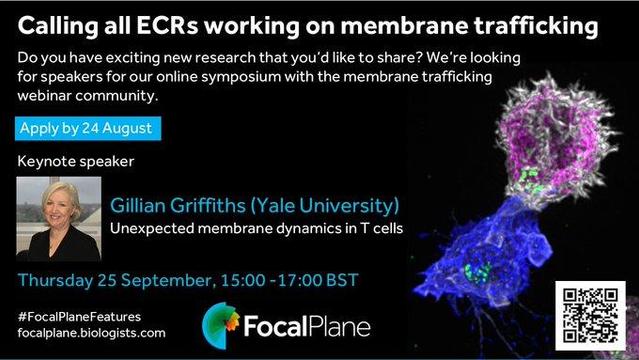
📢 Call for speakers
We are looking for early-career researchers to speak in FocalPlane's mini-symposium run together with the popular membrane trafficking webinar series organised by Francesca Bottanelli, Fèlix Campelo and Ishier Raote.
Application deadline: Sunday 24 August
Find out more:
#FocalPlaneFeatures #Microscopy #MembraneTrafficking #Cells #TCells #ECR #ImmunologicalSynapse #MembraneDynamics #Microscope #CellBiology
