#Methode
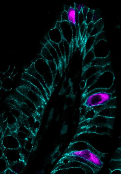
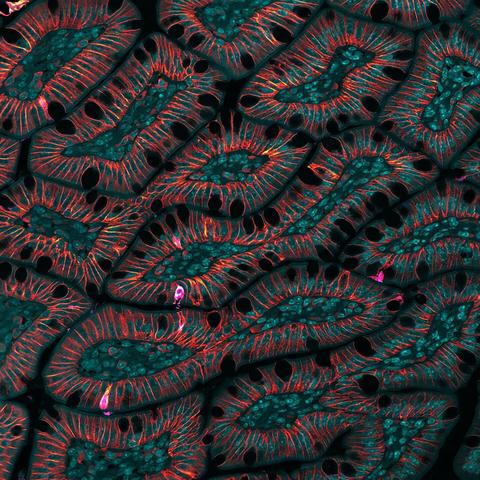
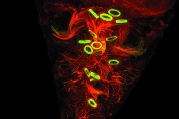
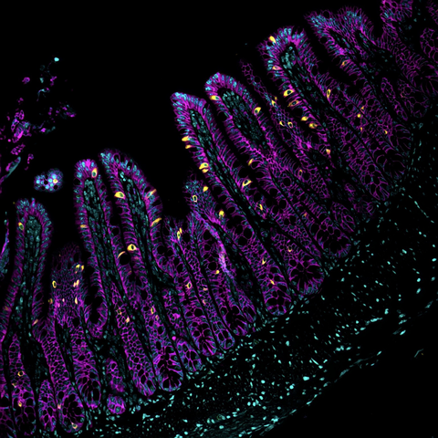
Fluoreszenz-Marathonläufer vs. Sprinter: Forschende der @unigoettingen
nutzen #FluoreszenzlebensdauerMikroskopie & HaloTag-Nanobodies, um Zielproteine zu detektieren und trotz spektraler Überlappung exakt auseinander zu halten.

Mehr im Artikel von Andrea Pitzschke: https://www.laborjournal.de/editorials/3450.php

#Laborjournal #LifeSci #FLIM #Nanobodies #HaloTag #Mikroskopie #Zellbiologie #MultiplexImaging #Immunofluorescence

The IBEX Knowledge-Base is setting a new standard for reproducibility in #immunofluorescence imaging: open, community-driven, and with ‘exceptional’ evidence of its utility.

It’s now up to the community to contribute to its growth.

🔗 https://elifesciences.org/reviewed-preprints/105737?utm_source=mastodon&utm_medium=social&utm_campaign=organic

This is an #immunofluorescence image of a section of mouse testis spermatocytes, Sertoli cells, and GPX4. Researchers investigated the impact of mitochondrial defects on sperm cells. #Mitochondria #12DaysOfeLife https://elifesciences.org/articles/84710?utm_source=mastodon&utm_medium=social&utm_campaign=organic_12Days
Mitochondrial defects caused by PARL deficiency lead to arrested spermatogenesis and ferroptosis

Mitochondrial functional and structural defects caused by PARL deficiency lead to arrested spermatogenesis and germ cell ferroptosis.

eLife

@domhenri Wow, those numbers are depressing.

Figure 3 should scare everyone who uses antibodies (and hopefully spur them to sanity-check their stainings with additional techniques).

https://elifesciences.org/reviewed-preprints/91645v1#fig3

#bioscience #immunostaining #immunofluorescence

Scaling of an antibody validation procedure enables quantification of antibody performance in major research applications

New concepts for #spatialbiology combining #immunofluorescence and #histology for next-gen #tissue analyses. Here, #machinelearning is used for multi-plexed
Immunofluorescence imaging and classical #histology of the same tissue section. With it a showcase illustrates for combined models of immune infiltration and tumor-intrinsic features a clinical read-out: a 10- to 20-fold improved discrimination between rapid and slow (or no) progression in #colorectalcancer.

https://www.nature.com/articles/s43018-023-00576-1

High-plex immunofluorescence imaging and traditional histology of the same tissue section for discovering image-based biomarkers - Nature Cancer

Sorger and colleagues present Orion, a technique to analyze H&E histology and multiplex immunofluorescence imaging data from the same cells, which they apply to human colorectal cancer samples to identify spatial biomarkers of disease progression.

Nature

Wow!
looks like super awesome method from Long Cai lab

"High-resolution spatial multi-omics reveals cell-type specific nuclear compartments"

They did mRNA #seqFISH+ (1200 genes, 17K introns), DNA seqFISH+ (100K loci) and #immunofluorescence (up to 65 antibodies) all in the same experiment!
200+ rounds of #imaging! That's incredible.

#mRNA #mRNALocalization #smFISH #DNA #3D_genome
#transcription #microscopy

https://www.biorxiv.org/content/10.1101/2023.05.07.539762v1

Dclk1+ cells in the intestinal epithelium 🔬 imaged by #immunofluorescence #microscopy
Impaired growth factor signaling alters the cellular differentiation pattern of the small intestine #immunofluorescence #microscopy

Fluorescein isothiocyanate (FITC) is an organic fluorescent dye and probably one of the most commonly used in #immunofluorescence and flow cytometry.
Learn more about Fluorescent Dyes: https://fcld.ly/b5zd1oh

🖼️ Drosophila embryo development, Green: FITC, Red: TRITC.

#fluorescencefriday #fluorescence #dyes #microscopy #science #sciencemastodon

Fluorescent Dyes

A basic principle in fluorescence microscopy is the highly specific visualization of cellular components with the help of a fluorescent agent. This can be a fluorescent protein – for example GFP – genetically linked to the protein of interest. If cloning is impossible – for instance in histologic samples – techniques such as immunofluorescence staining are used to visualize the protein of interest.

Tuft cells speckling the small intestine

#sciart #immunofluorescence #microscopy