Woman has #cyst removed and is shocked to learn it had #teeth, hair, and maybe even an #eyeball
https://www.upworthy.com/womans-cyst-has-teeth-hair-eyeball-ex1
Woman has #cyst removed and is shocked to learn it had #teeth, hair, and maybe even an #eyeball
https://www.upworthy.com/womans-cyst-has-teeth-hair-eyeball-ex1
Getting that huge old cyst drained is nasty business but I’m happy I did.
Pretty sure it’s plotting vengeance but I should be under a surgeon’s knife before it can muster any.
Still sleeping on a towel in bed but can probably stop.
If you ever get a plugged pore, take care of it early!
I was about ready to dress as Baron Harkonen. 😄

Scientists elucidate #molecular mechanisms behind #dinoflagellate #cyst #dormancy.
#cDNA #DinoSL #genomics #metatranscriptome #hormones #ABA #GA #metabolomics
https://phys.org/news/2025-02-scientists-elucidate-molecular-mechanisms-dinoflagellate.html

Dinoflagellates play crucial roles in aquatic ecosystems, particularly as major contributors to harmful algal blooms. They can enter a dormant stage, known as the resting cyst stage, that allows them to survive for extended periods—up to 150 years—in marine sediments. This dormancy is essential for their annual population dynamics, blooming cycles, and geographic expansion.
Our #preprint where we derive an #activeGel #model with entropic elasticity of the #microstructure from the thermodynamic constraints on the dynamics of #myosin molecular motors is now updated!
Hopefully more readable, and with the example of a #cyst like contractile sphere.
#cytoskeleton #rheology #activeMatter #softMatter #actomyosin
god I can't wait for this election to be over
ovary: you know what would be worse?
...please no
ovary: if i blew a couple of cysts right now
whyyyyy
#pcos #cyst #ovariancyst #ovaries #ohno #myovaries #womenshealth
The ultrasound technician said it looks like a cyst. Now waiting for the X-ray.
Fucking cysts!
At least there’s little chance a finger cyst will grow to be the size of a large grapefruit like an ovarian cyst. Also, pelvic ultrasounds are *the worst* because of the need to drink a LOT of water beforehand while not being allowed to pee until after they press down on your full bladder to see the fucking cyst. Talk about torture.
Diffusion MRI shows potential in #cyst and tissue assessment in autosomal dominant polycystic #kidney #disease.
#EuropeanRadiology #Fibrosis #Radiology
Read the full article now ➡️ https://link.springer.com/article/10.1007/s00330-023-09601-4
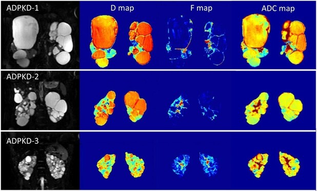

Objectives Beyond total kidney and cyst volume (TCV), non-cystic tissue plays an important role in autosomal dominant polycystic kidney disease (ADPKD) progression. This study aims at presenting and preliminarily validating a diffusion MRI (DWI)–based TCV quantification method and providing evidence of DWI potential in characterising non-cystic tissue microstructure. Methods T2-weighted MRI and DWI scans (b = 0, 15, 50, 100, 200, 350, 500, 700, 1000; 3 directions) were acquired from 35 ADPKD patients with CKD stage 1 to 3a and 15 healthy volunteers on a 1.5 T scanner. ADPKD classification was performed using the Mayo model. DWI scans were processed by mono- and segmented bi-exponential models. TCV was quantified on T2-weighted MRI by the reference semi-automatic method and automatically computed by thresholding the pure diffusivity (D) histogram. The agreement between reference and DWI-based TCV values and the differences in DWI-based parameters between healthy and ADPKD tissue components were assessed. Results There was strong correlation between DWI-based and reference TCV (rho = 0.994, p < 0.001). Non-cystic ADPKD tissue had significantly higher D, and lower pseudo-diffusion and flowing fraction than healthy tissue (p < 0.001). Moreover, apparent diffusion coefficient and D values significantly differed by Mayo imaging class, both in the whole kidney (Wilcoxon p = 0.007 and p = 0.004) and non-cystic tissue (p = 0.024 and p = 0.007). Conclusions DWI shows potential in ADPKD to quantify TCV and characterise non-cystic kidney tissue microstructure, indicating the presence of microcysts and peritubular interstitial fibrosis. DWI could complement existing biomarkers for non-invasively staging, monitoring, and predicting ADPKD progression and evaluating the impact of novel therapies, possibly targeting damaged non-cystic tissue besides cyst expansion. Clinical relevance statement This study shows diffusion-weighted MRI (DWI) potential to quantify total cyst volume and characterise non-cystic kidney tissue microstructure in ADPKD. DWI could complement existing biomarkers for non-invasively staging, monitoring, and predicting ADPKD progression and evaluating the impact of novel therapies, possibly targeting damaged non-cystic tissue besides cyst expansion. Key Points • Diffusion magnetic resonance imaging shows potential to quantify total cyst volume in ADPKD. • Diffusion magnetic resonance imaging might allow to non-invasively characterise non-cystic kidney tissue microstructure. • Diffusion magnetic resonance imaging–based biomarkers significantly differ by Mayo imaging class, suggesting their possible prognostic value.

