Cellule nervose al microscopio ottico

In blu scuro troviamo il corpo centrale dei neuroni, chiamato pirenoforo.

Da questo si diramano tanti piccoli filamenti, i dendriti, che servono per la ricezione degli impulsi, e l'assone, un prolungamento che serve per la trasmissione.

I puntini blu che circondano i neuroni sono i nuclei delle cellule gliali, il cui compito è quello di sostenere e aiutare i neuroni.

Riuscite a vederle?

#nature #histology #microscopyimages

Quartz crystal fun w Affinity Photo stacking software, thanks to Anglian Microscopy Group photo tuition yesterday and Quekett Microscopical Club funding + Stephanie @ Art Branches for vuggy Somerset specimen.
A first attempt, using #Motorola E 'phone 📷 w Slokey adapter on Meiji stereo microscope.
Magn. c.10x, FOV c.10 mm.
#geology #minerals #microscopyimages #MicroscopyMonday
#AcademicMastodon is ¿trending? so I guess I should write an #introduction. I'm a clinician scientist / #pathologist at the Institute of Cancer Research #london, specializing in #uropathology and #ImageAnalysis #AI. Quite into US, UK and Turkish #politics and #economics as well. Interested in #running #scuba #astronomy and #astrophysics , as well as #sciencefiction and #gaming . Follow me for #pathology tidbits and cool #microscopyimages

Microscopic Photo of an Oak Leaf Wins Nikon’s Small World Competition

Nikon has announced the winners of its 47th annual Small World Photomicrography Competition. This year’s first-place prize was awarded to Jason Kirk for his striking image of a southern live oak leaf’s trichomes, stomata, and vessels.

The Nikon Small World competition was founded in 1974 to recognize excellence in photography through the microscope and is widely regarded as the leading forum for recognizing the art, proficiency, and photographic excellence involved in photomicrography. In 2011, the sister competition of Nikon Small World, Nikon Small World in Motion, was launched in response to technology advances allowing for recording movies or digital time-lapse photography through the microscope. The winners of that competition were announced on August 16.

This year, the competition received nearly 1,900 entries from 88 countries which were judged on originality, informational content, technical proficiency, and visual impact.

To capture his photo of an oak leaf, Kirk used various lighting techniques and design tools to create what Nikon's judges determined to be a masterful example of the dynamic relationship between imaging technology and artistic creativity. Using a custom-made microscope system that combines color-filtered transmitted light with diffused reflected light, Jason captured around 200 individual images of the leaf and stacked them together to create the stunning image above.

Jason used both transmitted and reflected light on opposite sides of the leaf to highlight three vital structures. Prominently featured in white are the trichomes, which are fine outgrowths that protect a plant against extreme weather, microorganisms, and insects. In purple, Jason highlights the stomata, small pores that regulate the flow of gases in a plant. Colored in cyan are the vessels that transport water throughout the leaf. All three are essential to plant life.

Second place was awarded to Esmeralda Paric for her image of a microfluidic device containing hundreds of thousands of networking neurons. The primary neurons were extracted and cultured, then seeded and transduced with a virus. The particular image shows two populations separated but bridged, with different viral treatments. It was maintained for 30 days, immunostained, and tiled imaged.

A microfluidic device containing 300k networking neurons in 2 isolated populations. Both sides were treated with a unique virus and bridged by axons. | Esmeralda Paric & Holly Stefen

Third place went to Frank Reiser for his photo of a rear leg, claw, and respiratory trachea of a hog louse (Haematopinus suis).

Rear leg, claw, and respiratory trachea of a louse (Haematopinus suis) | Frank Reiser

Below are the 17 other images -- in order from fourth through 20th place -- recognized in the top 20 photos submitted to the competition:

Sensory neuron from an embryonic rat | Paula Diaz Slime mold (Arcyria pomiformis) | Alison Pollack Calcite crystal inclusion suspended in a spinel gemstone | Billie Hughes Table salt crystal | Saulius Gugis Filamentous strands of Nostoc cyanobacteria captured inside a gelatinous matrix | Martin Kaae Kristiansen An in vivo snapshot of the neurons surrounding the mouth and tentacles of a juvenile starlet sea anemone (Nematostella vectensis) | Ruohan Zhong Diatom (Arachnoidiscus) | Bernard Allard Snowflake | Dr. Joern N. Hopke Cotton fabric with pollen grains | Dr. Felice Placenti Breast organoid showing contractile myoepithelial cells (blue) crawling on secretory breast cells (red) | Jakub Sumbal Vasculature of a mouse retina | Jason Kirk & Carlos P. Flores Suarez Vein and scales on a butterfly wing (Morpho didius) | Sébastien Malo Water flea (Daphnia), carrying embryos and peritrichs | Jan van IJken Cross section of mouse intestine | Dr. Amy Engevik Head of a tick | Dr. Tong Zhang & Dr. Paul Stoodley 3D vasculature of an adult mouse brain (somatosensory cortex) | Dr. Andrea Tedeschi Proboscis of a housefly (Musca domestica) | Oliver Dum

A full-color calendar of all the winners is planned to be produced. To see all images from the competition that were recognized by the judges, visit the Nikon Small Worlds Competition website.

Image credits: Header image by Jason Kirk. All other photos individually credited and provided courtesy of Nikon Small World.

#features #inspiration #news #beautiful #colorful #gorgeous #incredible #microscope #microscopic #microscopicphotography #microscopy #microscopyimages #microscopyphotos #nikon #nikonsmallworld #nikonsmallworld2021 #stacking #stunning

Microscopic Photo of an Oak Leaf Wins Nikon's Small World Competition

The beauty in the small.

Olympus Unveils the Winners of the Best Life Science Photos of 2020

Olympus has announced the winners of its Global Image of the Year Life Science Light Microscopy Award, which is an annual competition that recognizes the best in life science photos taken with a microscope.

This competition is hosted by Olympus's Life Sciences division, which is not to be confused with the newly-formed OM Digital under Japan Industrial Partners. The competition started in 2017 as the Image of the Year European Life Science Light Microscopy Award with the aim to celebrate both the artistic and scientific value of microscopy images. Today, the organizers say that the competition stays true to this mission by encouraging people across the world to look at scientific images in a new way, appreciate their beauty, and share images with others.

To be considered for this year's competition, images had to be submitted between September 15, 2020 and January 31, 2021. A maximum of three images was allowed per participant, and anyone over the age of 18 could participate (provided that they are not an Olympus employee, family member, judge, or family member of a judge nor engaged in the fields of manufacturing or sales of microscopes).

All entries were evaluated on artistic and visual aspects, scientific impact, and microscope proficiency.

From rat embryos to butterfly scales and snakeskin, the Olympus Life Science team was impressed by the diverse collection captured under the microscope this year.

This year's competition saw nearly 700 submissions from 61 different countries, with the winning image going to Werner Zuschratter from Germany. Zuschratter submitted the incredible photo below depicting a whole rat embryo that he captured with a confocal microscope. For the grand prize, Zuschratter will receive an Olympus SZX7 stereo microscope with a DP27 digital camera.

In addition to the overall global winner, three regional awards were presented to XinPei Zhang (China) for Asia, Justin Zoll (USA) for the Americas, and Grigorii Timin (Switzerland) for EMEA. Each regional winner will receive an Olympus CX23 upright microscope.

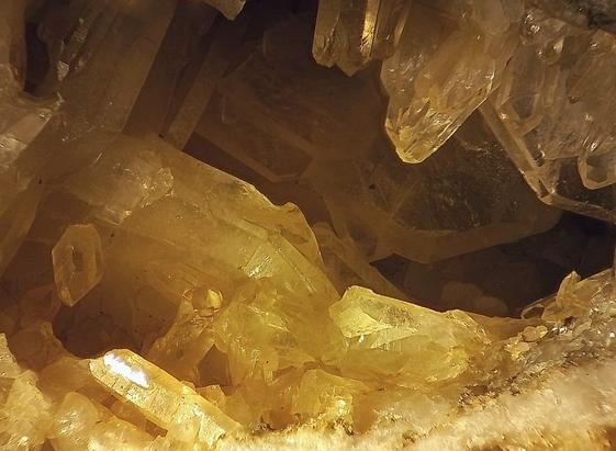
Collagen fibers (second harmonic generation) and dermal pigment cells (autofluorescence) in African house snake embryonic skin; maximal intensity projection of 10 confocal slices. | Regional Winner - EMEA - Grigorii Timin - Switzerland Scales collected from the wings of over 40 species of butterflies were photographed individually and finally assembled into this image. | Regional Winner - Asia Pacific - XinPei Zhang - China Polarized light microscopy panorama of L-glutamine and beta alanine crystals | Regional Winner - Americas - Justin Zoll - United States

Though they may not have brought home the grand prize or won in any of the three regions, the honorable mentions below are still breathtaking images.

Mouse testes stained for Sertoli cells (blue and red) | Derek Sung, United States Anther of Arabidopsis arenosa stained with aniline blue, Z-stack of confocal sections (maximum intensity projection) | Jan Martinek, Czech Republic Whole-mount immunofluorescence showing the muscles (cyan, F-actin) and the nervous system (yellows, acetylated tubulin) counterstained by the nuclei (blue, DNA) of a clarified and decalcified post-metamorphic sea urchin juvenile (Paracentrotus lividus). | Laurent Formery, France A polarized light micrograph of crystals formed by the rapid evaporation of water from an aqueous solution of Beta-Alanine (a popular training supplement), and L-glutamine (an essential amino acid). Home-made tunable wave plate. | Matt Inman, Australia Microfilament structure in U2OS labeled with fluorescent protein, 3D projection image acquired by airyscan | MingShu Zhang, China Primary rat cortical neurons at 14 days in vitro with nuclei (green) and developing dendrites (mpl-inferno LUT) | Nadia Efimova, United States
The sample shows a dividing human embryonic kidney (HEK) 293 cell. Green marks the cellular boundary, red marks the mitochondria, while blue shows the separating chromosomes. | Sayantan Datta, India Wing scales of the twilight moth Urania ripheus. | Walter Ferrari, Argentina “Pollock’s glia”. 3D reconstructed immunostaining image of astrocyte (GFAP+, white), oligodendrocyte (NG2+, blue), and microglia (IBA1+, red) in the brain white matter, which is highly similar to Pollock’s Abstract paintings. | YiXun Su, China

To read about the jurors and see past competition winners, visit the Olympus Lifescience website here.

Image credits: All photos individually credited and provided courtesy of Olympus Life Science.

#features #finds #lifescience #lifescienceimages #lifesciencephotos #microscope #microscopy #microscopyimages #microscopyphotos #olympuslifesciences

Olympus Unveils the Winners of the Best Life Science Photos of 2020

Under the microscope.