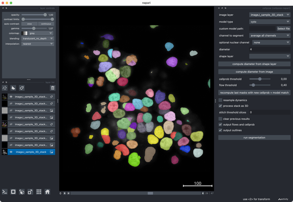
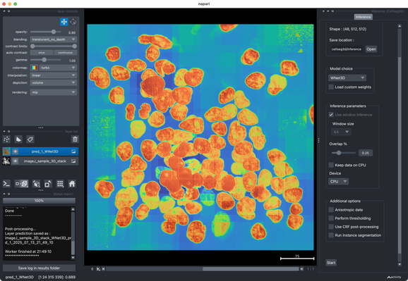
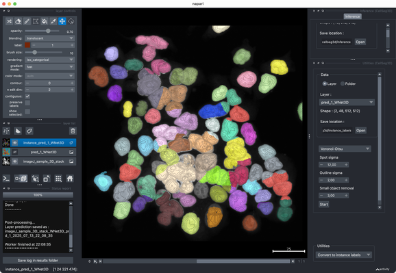
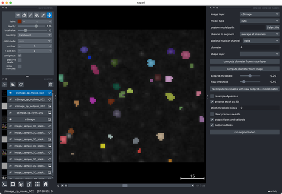
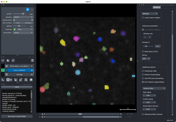
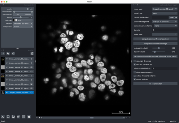
Tried the same with a more realistic 3D stack from the #ImageJ sample library.#Cellpose runs fast and segments very well out of the box.#CellSeg3D takes considerably longer and seems to segment decently, but I couldn’t get a proper instance #segmentation in the post-processing step (which is recommended as part of its workflow). However, #CellSeg3D looks very promising — just needs some more time and parameter exploration, I guess.

I’d recommend giving it a try 👌
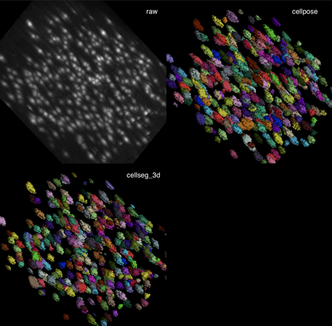
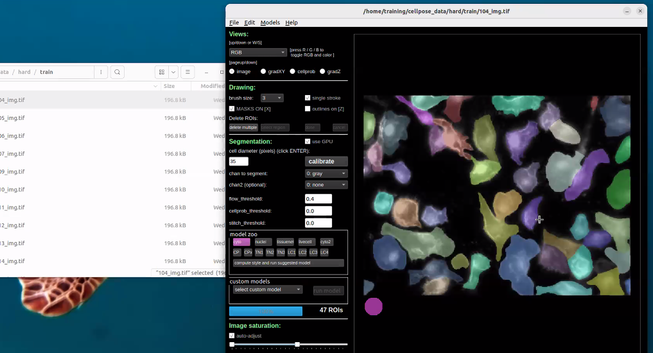
Tested #CellSeg3D and #Cellpose on their example c5image dataset. Both segmentations look reasonable out-of-the-box, without any deep parameter tuning. With some extra effort, one could likely push either further I guess. Overall, both tools perform quite well on this small sample data set.

✍️ New in #eLife: #CellSeg3D introduces #WNet3D, a self-supervised 3D #segmentation method for #microscopy data — no labels needed. Claims to outperform #Cellpose/#StarDist on 4 datasets. Includes #opensource plugin (#Napari) + full 3D annotated #cortex dataset. Will test it later.

🌍 https://elifesciences.org/articles/99848

#DeepLearning #Neuroscience

CellSeg3D, Self-supervised 3D cell segmentation for fluorescence microscopy

Self-supervised deep learning models can accurately perform 3D segmentation of cell nuclei in complex biological tissues, enabling scalable analysis in settings with limited or no ground truth annotations.

eLife

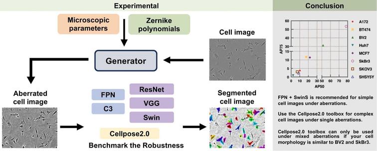
🤖 How resilient are deep learning models to real-world imperfections in microscopy?

🔗 Practical guidelines for cell segmentation models under optical aberrations in microscopy. Computational and Structural Biotechnology Journal, DOI: https://doi.org/10.1016/j.csbj.2024.09.002

📚 CSBJ Quantum Biology and Biophotonics: https://www.csbj.org/qbio

#CellSegmentation #Microscopy #AIinBiology #OpticalAberrations #DeepLearning #BiomedicalAI #Cellpose #ComputationalBiology #PLCM

After a day and a half of #ImageAnalysis in the cloud with #dask by the IDR team, Damian Dalle Nogare takes over a practical session to apply several #AI models for cell segmentation using #Cellpose

Despite all their hype in AI, we found that Transformers do not outperform #Cellpose for cellular segmentation tasks: https://www.biorxiv.org/content/10.1101/2024.04.06.587952v1

#bioinformatics #biology #machinelearning

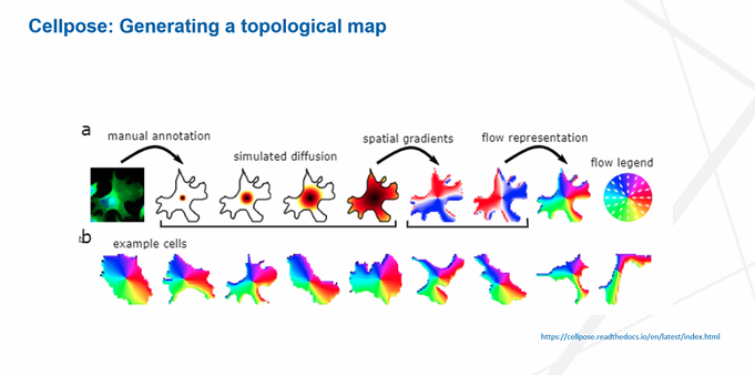
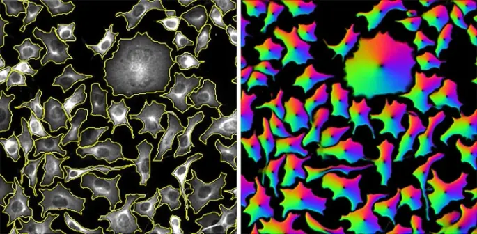
#Cellpose 3! Not all images are perfect. Restore your images with Cellpose3 to get better segmentations, w/ @marius10p https://www.biorxiv.org/content/10.1101/2024.02.10.579780v1

(click to play gif)

AI under the microscope: the algorithms powering the search for cells: https://go.nature.com/3SVdFur. Nice article by Michael Eisenstein about cellular segmentation, with a shout out to #cellpose :) #biology #deeplearning
AI under the microscope: the algorithms powering the search for cells

Deep learning is driving the rapid evolution of algorithms that can automatically find and trace cells in a wide range of microscopy experiments.

@ogi thanks, I did not realize this before, that's great. and yes we use #pyqtgraph in a bunch of projects, love how fast and flexible it is! #cellpose is our most popular software tool that uses it: https://github.com/MouseLand/cellpose
GitHub - MouseLand/cellpose: a generalist algorithm for cellular segmentation with human-in-the-loop capabilities

a generalist algorithm for cellular segmentation with human-in-the-loop capabilities - MouseLand/cellpose

GitHub

Cell/nuclei segmentation by training deep learning models for #CellPose and #StarDist incrementally, using sparse annotations

Latest work by Ko Sugawara, initiated in our team and completed at RIKEN

https://www.biorxiv.org/content/10.1101/2023.06.13.544786v1.full.pdf