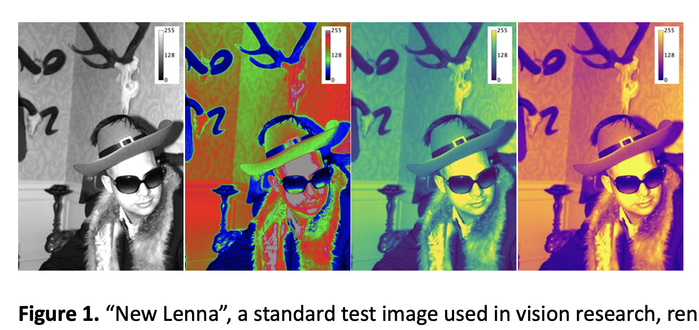
Ah, feeling nostalgic, my former colleague became the new poster child for image tests in computer vision: a test image built in #ImageJ # FIJI
(I am making figure to explain perceptionally uniform #color scales...)
#NewLenna #Lenna #DataVis
Ah, feeling nostalgic, my former colleague became the new poster child for image tests in computer vision: a test image built in #ImageJ # FIJI
(I am making figure to explain perceptionally uniform #color scales...)
#NewLenna #Lenna #DataVis
Fighting #ImageJ today. It's losing greyscale calibration when I crop a section from a larger image.
That said in typing this I think I worked out how to fix it. Thank you all for being my rubberduck.
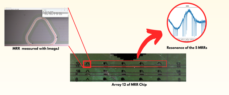
How would a signal look like from our micro-ring resonator #MRR #biosensor?
We observe a spectrum in a wavelength range around 1.55 µm by coupling the laser radiation into an array of five MRRs, see bottom figure. It is notable that each MRR has a curved triangular shape of different size this causes the resonance peak of each MRR to occur at different positions in the spectrum.
In the spectrum range investigated, we observed four groups of resonance peaks corresponding to four standing waves occurring to each of the five rings.
Determining the perimeter of the ring was challenging. I used #ImageJ to draw a polygon that approximated the triangular shape and measured the perimeter. Then, with the use of the formulas obtained from P. Steglich, et al(https://ieeexplore.ieee.org/abstract/document/9568878), it was possible to determine the effective refractive index nff and the order m of the standing wave.
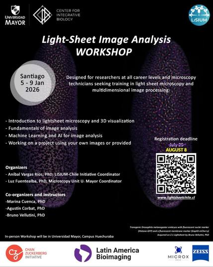
Light-Sheet Image Analysis Workshop 2026
The Light-Sheet Image Analysis Workshop is a five-day intensive course that will take place in Santiago, Chile, from January 5–9, 2026, designed for students and researchers who wish to gain foundational skills in the processing and analysis of light-sheet microscopy imaging data.
The application deadline has been extended until August 8, 2025! The workshop is free to attend and travel fellowships are available. Learn more and apply here:
https://lightsheetchile.cl/light-sheet-image-analysis-workshop-2026-2/
—
URL: https://brunovellutini.com/posts/light-sheet-image-analysis-workshop-2026/
This is great spatially-resolved transcriptomics by the R/Bioconductor package visiumStitched, which facilitates stitching the images together with Fiji (ImageJ). 🦾
Bin heute mit den Automatismen von #ImageJ / #Fiji nicht vollends zufrieden gewesen. Wahrscheinlich sind meine Partikel zu klein.
Jetzt habe ich das manuell erledigt.
Hier lohnt sich mal wieder die Stifteingabe.
Habe insgesamt über 300 Partikel analysiert, das genügt für die grobe Statistik und ich kann zeigen, dass die so grob 10 nm Durchmesser haben.
#MöphDiss
Edit: Das zweite Bild ist nur ein Zwischenstand.
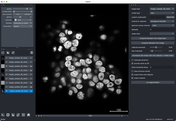
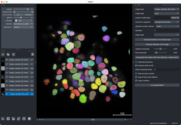
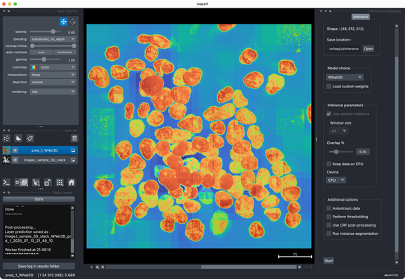
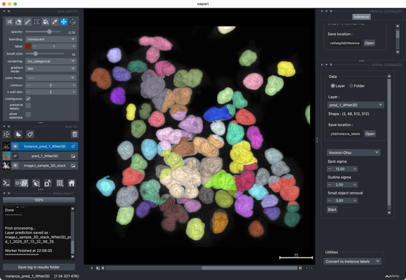
Tried the same with a more realistic 3D stack from the #ImageJ sample library.#Cellpose runs fast and segments very well out of the box.#CellSeg3D takes considerably longer and seems to segment decently, but I couldn’t get a proper instance #segmentation in the post-processing step (which is recommended as part of its workflow). However, #CellSeg3D looks very promising — just needs some more time and parameter exploration, I guess.
I’d recommend giving it a try 👌
I promised myself a while back that I wouldn't write any more #ImageJ Macro code. When I made this promise I forgot about all the scripts that we have in use. “Oh, this one just needs a few tweaks and then it can be used in another project." And here I am again in ijm hell!