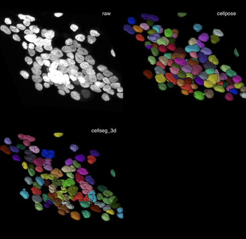
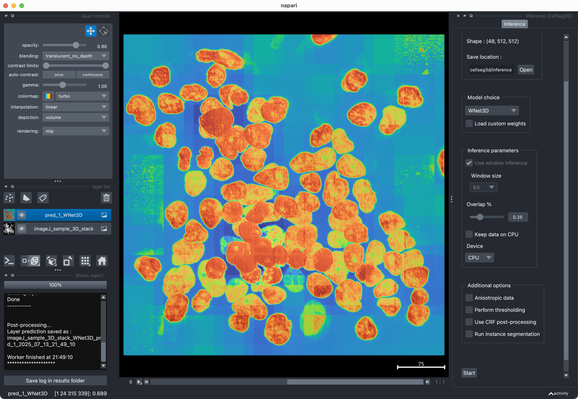
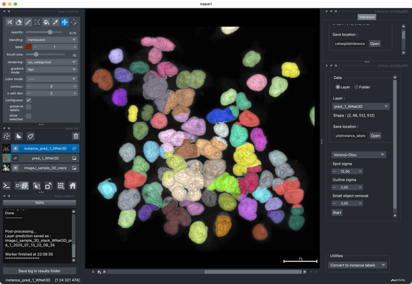
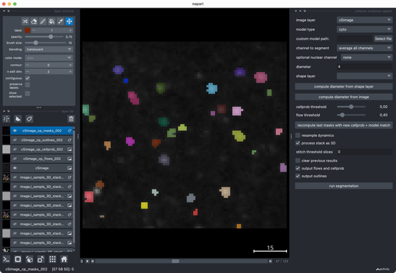
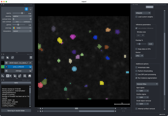
To wrap this up: Both tools are easy to test. I highly recommend trying them on your own data to see what works best for your use case.

Iโ€™ll include #CellSeg3D in our next #Napari #bioimage analysis course (https://www.fabriziomusacchio.com/teaching/teaching_bioimage_analysis/). Curious what impressions and feedback the students will share. ๐Ÿงช๐Ÿ”

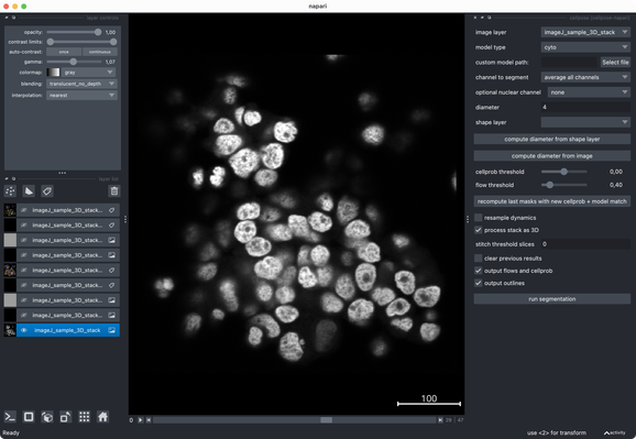
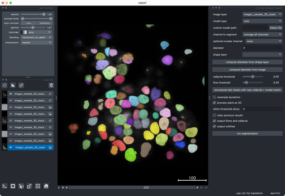
What I really like about @napari is how well it integrates modern #Python tools. Great to have such a flexible, evolving #opensource platform for (bio) #imageanalysis! ๐Ÿ‘Œ

Tried the same with a more realistic 3D stack from the #ImageJ sample library.#Cellpose runs fast and segments very well out of the box.#CellSeg3D takes considerably longer and seems to segment decently, but I couldnโ€™t get a proper instance #segmentation in the post-processing step (which is recommended as part of its workflow). However, #CellSeg3D looks very promising โ€” just needs some more time and parameter exploration, I guess.

Iโ€™d recommend giving it a try ๐Ÿ‘Œ

Tested #CellSeg3D and #Cellpose on their example c5image dataset. Both segmentations look reasonable out-of-the-box, without any deep parameter tuning. With some extra effort, one could likely push either further I guess. Overall, both tools perform quite well on this small sample data set.

โœ๏ธ New in #eLife: #CellSeg3D introduces #WNet3D, a self-supervised 3D #segmentation method for #microscopy data โ€” no labels needed. Claims to outperform #Cellpose/#StarDist on 4 datasets. Includes #opensource plugin (#Napari) + full 3D annotated #cortex dataset. Will test it later.

๐ŸŒ https://elifesciences.org/articles/99848

#DeepLearning #Neuroscience

CellSeg3D, Self-supervised 3D cell segmentation for fluorescence microscopy

Self-supervised deep learning models can accurately perform 3D segmentation of cell nuclei in complex biological tissues, enabling scalable analysis in settings with limited or no ground truth annotations.

eLife