I am not sure what this is. I am not sure what anything is.
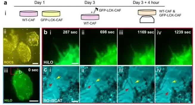
🔬 A breakthrough for spatial biology? This ultrafast 3D imaging method enables deep whole-organ visualization faster than ever before.
🔗 Rapid 3D Immunolabeling and Light Sheet Microscopy for Quantitative Analysis of Intact Tissues. Computational and Structural Biotechnology Journal (CSBJ). DOI: https://doi.org/10.34133/csbj.0121
📚 CSBJ - A Science Partner Journal: https://spj.science.org/journal/csbj
#Bioimaging #3DImaging #LightSheetMicroscopy #TissueBiology #Microscopy #CellBiology #SpatialBiology #Histology

Breakthrough Imaging Technique Exposes Elusive Cell-to-Cell Networks
Australian National University develops new microscope to see how cells talk to each other in 3D. This helps understand diseases and how viruses spread.
#CellCommunication, #ANU, #Microscopy, #DiseaseResearch, #ViralSpread
https://newsletter.tf/anu-nanoscopy-sees-hidden-cell-networks/
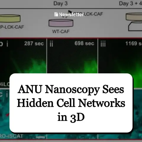
Scientists can now see tiny cell connections in 3D for days, like watching a hidden city map of cell messages. This is a big step from older microscopes.
#CellCommunication, #ANU, #Microscopy, #DiseaseResearch, #ViralSpread
https://newsletter.tf/anu-nanoscopy-sees-hidden-cell-networks/
Open position: Systems Engineer II.
#science #engineering #career #jobs #job #jobsearch
#physics #imaging #microscopy
www.lifescience.net/jobs/230191/...
Systems Engineer II
Systems Engineer II
Well it’s the final day of what has been pretty much my ideal meeting! JCS Imaging Cell Dynamics has been an absolute blast. Fantastic organisation by @Co_Biologists in an amazing venue in the hills of Catalonia. #microscopy #cellbiology

Glass slide 🇺🇸
#microscope #microscopist #microscopy #sample
▶️ 1 new picture from NIH BioArt https://commons.wikimedia.org/wiki/File:Glass_slide_%28NIH_BioArt_679%29.svg

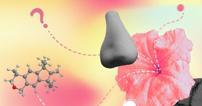
👃🧬 Scientists are using cryo-electron #microscopy to visualize how scent #molecules bind to receptors in the nose.
The structural details help explain how the #brain distinguishes between thousands of different odors based on molecular shape and #chemistry. The findings provide a clearer understanding of the biological process that allows us to experience the world through smell.
👉 https://www.chemistryworld.com/features/the-molecular-mystery-of-how-we-smell/4023405.article
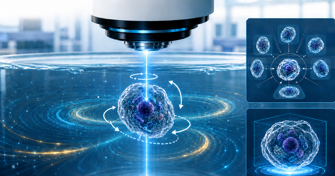
A non-contact technique that utilizes laser-induced thermo-viscous fluid flows to rotate delicate microscopic samples in all three spatial dimensions.
#Microscopy #OpticalImaging #FluidDynamics #Biophysics #Microrobotics #Microtechnology #sflorg
https://www.sflorg.com/2026/05/phy05122601.html
#Microscopy #OpticalImaging #FluidDynamics #Biophysics #Microrobotics #Microtechnology #sflorg
https://www.sflorg.com/2026/05/phy05122601.html
New Paper Day!
A hyper specific investigation of how Ameloblasts get incorporated into enamel as it forms, and what this does.
Enamel Tubules and Spindles: Enter and Exit the Amelocyte | Calcified Tissue International | Springer Nature Link
https://link.springer.com/article/10.1007/s00223-026-01522-w
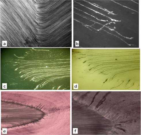
Enamel Tubules and Spindles: Enter and Exit the Amelocyte - Calcified Tissue International
By the commonly accepted rules of nomenclature, cells forming hard tissue which become incorporated within the inter- and extra-cellular tissue mass which they secrete are called ‘-cytes’: the spaces in which these cells sit are called lacunae. Hence, osteoblasts become osteocytes in osteocytic lacunae, chondroblasts become chondrocytes, cementoblasts cementocytes, even odontoblasts become odontocytes in some mammalian species. We provide evidence that enamel spindles are cell bodies of ameloblasts which have become entrapped and buried in the enamel matrix produced by the surrounding cells, to produce amelocytic lacunae: it is no life for a cell in a rigid, dense and dead tissue, and we do not suppose that amelocytes live once they are set on that route. Spindles, normally only found at the enamel-dentine junction, are absent when the enamel at the junction is free of prisms, i.e., the ameloblasts had no Tomes’ processes. Spindles have an organic content, the amelocyte residue which can be stained - and removed by bleach, after which the spindle space no longer stains. Spindles are extensions of enamel tubules which result from the extension of a fine ameloblastic process [from the major Tomes’ process] which unites with an odontoblast process. In marsupial mammals, the spindle-like ends of the enamel tubules are commonly located well within the enamel. Spindles/amelocytes may form at the neonatal line in human deciduous teeth in cases of severe neonatal stress, and these features simply represent encapsulation of moribund ameloblasts. The frequency of spindles at the enamel dentine junction is greater in cases where the tooth germ is severely deformed by growth pressure from the forming root of a neighbouring tooth.