Sometimes one stops to just look at the data. And the software user interface. They are beautiful.

We are looking at a cross section of the #Drosophila larval brain, near the brain commissure, where hundreds of neurons (magenta: their reconstructed skeletons) cross from one brain hemisphere to the other. To the right, a 3D rendering of multiple neurons, a pair of which cross the midline in a U-shaped bend.

Our CATMAID software is web-based, in other words it's just a website that accesses remote data. I credit it's sleek design to @herrsaalfeld – author of the early, "Ice Age" CATMAID and its blue tones – who at some point in his life studied "medieninformatik" and has always had a penchant for art.

See our images and fly neurons here, kindly hosted by the #VirtualFlyBrain :
https://l1em.catmaid.virtualflybrain.org/?pid=1&zp=33100&yp=31179&xp=53333&tool=tracingtool&sid0=1&s0=2

#neuroscience #CATMAID

CATMAID

@CurrentBiology

Andreas Schoofs and Anton Miroschnikow in Michael Pankratz' lab have done an astonishing amount of work manually mapping the peripheral nervous system of the larval #Drosophila, with #CATMAID, in the STEM volume we imaged and they named "Igor". Congrats on seeing this gargantuan project through!

The whole larval volume of "Igor", including all tissues, is available here, courtesy of the #OpenOrganelle project led by Aubrey Weigel @avweigel at #HHMIJanelia:
https://openorganelle.janelia.org/datasets/jrc_fly-larva-1

See it at 5x5x35 nm resolution in #neuroglancer: https://neuroglancer-demo.appspot.com/#!%7B%22dimensions%22:%7B%22x%22:%5B1e-9%2C%22m%22%5D%2C%22y%22:%5B1e-9%2C%22m%22%5D%2C%22z%22:%5B1e-9%2C%22m%22%5D%7D%2C%22position%22:%5B249600.5%2C79040.5%2C84280.5%5D%2C%22crossSectionOrientation%22:%5B0%2C1%2C0%2C0%5D%2C%22crossSectionScale%22:50%2C%22projectionOrientation%22:%5B0%2C1%2C0%2C0%5D%2C%22projectionScale%22:65536%2C%22layers%22:%5B%7B%22type%22:%22image%22%2C%22source%22:%22n5://s3://janelia-cosem-datasets/jrc_fly-larva-1/jrc_fly-larva-1.n5/em/tem-uint8%22%2C%22tab%22:%22rendering%22%2C%22opacity%22:1%2C%22blend%22:%22additive%22%2C%22shader%22:%22#uicontrol%20invlerp%20normalized%28range=%5B0%2C%20255%5D%2C%20window=%5B0%2C%20255%5D%29%5Cn#uicontrol%20vec3%20color%20color%28default=%5C%22white%5C%22%29%5Cnvoid%20main%28%29%7BemitRGB%28color%20%2A%20normalized%28%29%29%3B%7D%22%2C%22name%22:%22tem-uint8%22%7D%5D%2C%22selectedLayer%22:%7B%22visible%22:true%2C%22layer%22:%22tem-uint8%22%7D%2C%22crossSectionBackgroundColor%22:%22#000000%22%2C%22layout%22:%224panel%22%7D (control-minus and control-plus to zoom; mouse click to pan, scroll wheel to browse in Z).

Will appear at the #VirtualFlyBrain website soon as well.

There are many more peripheral nervous system components mapped, yet to be published.

This paper is open access:
"Serotonergic modulation of swallowing in a complete fly vagus nerve connectome", Schoofs et al. 2024 https://www.cell.com/current-biology/fulltext/S0960-9822(24)01137-0?rss=yes&utm_source=dlvr.it&utm_medium=mastodon

#neuroscience #connectomics

Today the peer-reviewed version of our preprint is out:

"The #connectome of an insect brain"
https://www.science.org/doi/10.1126/science.add9330

Congrats to co-first authors Michael Winding and Benjamin Pedigo, and to all our lab members and collaborators who made this work possible over the years. A journey that started over 10 years ago–and yet this is but a new beginning. So much more to come.

See my #tootprint on the preprint from back in the Autumn: https://mathstodon.xyz/@albertcardona/109422190525090990

The data is available both as supplements and directly via #CATMAID thanks to hosting by the #VirtualFlyBrain:
https://l1em.catmaid.virtualflybrain.org/?pid=1&zp=108250&yp=82961.59999999999&xp=54210.799999999996&tool=tracingtool&sid0=1&s0=2.4999999999999996&help=true&layout=h(XY,%20%7B%20type:%20%22neuron-search%22,%20id:%20%22neuron-search-1%22,%20options:%20%7B%22annotation-name%22:%20%22papers%22%7D%7D,%200.6)

(The "Winding, Pedigo et al. 2023" annotation listing all included neurons will appear very soon in an upcoming update.)

#neuroscience #connectomics #Drosophila #DrosophilaLarva

@dantracey @kristinmbranson @annikabarber @debivort @giorgiogilestro

Find the original basin-1 neurons at the #VirtualFlyBrain at "Tools - CATMAID - Hosted EM Data - Larval - Larva (ABD1.5)" which opens a #CATMAID server https://abd1.5.catmaid.virtualflybrain.org/?pid=1&zp=10485&yp=40560.65722061269&xp=42396.0789533435&tool=tracingtool&sid0=1&s0=4.5&help=true&layout=h(XY,%20%7B%20type:%20%22neuron-search%22,%20id:%20%22neuron-search-1%22,%20options:%20%7B%22annotation-name%22:%20%22papers%22%7D%7D,%200.6)

Find them via Neuron Search (icon with a "?").

The "Construction time" is wrong (see "Summary info" of the Selection Table) because these neurons were imported from #TrakEM2. Old enough to predate the #CATMAID software!
#neuroscience

CATMAID

The web-based open source software #CATMAID was devised as "google maps but for volumes". Documentation at https://catmaid.org and source code at https://github.com/catmaid/CATMAID/

Modern #CATMAID enables hundreds of #neuroscience researchers world wide to collaboratively map neuronal circuits in large datasets limited only by bandwidth and server-side storage to map and analyse a whole brain #connectome.

See the #Drosophila larval CNS at the #VirtualFlyBrain server: https://l1em.catmaid.virtualflybrain.org/?pid=1&zp=108250&yp=82961.59999999999&xp=54210.799999999996&tool=tracingtool&sid0=1&s0=2.4999999999999996&help=true&layout=h(XY,%20%7B%20type:%20%22neuron-search%22,%20id:%20%22neuron-search-1%22,%20options:%20%7B%22annotation-name%22:%20%22papers%22%7D%7D,%200.6)

Welcome to CATMAID — CATMAID 2021.12.21 documentation

The Collaborative Annotation Toolkit for Massive Amounts of Image Data. Used for image viewing, neuron reconstruction, ontology based annotations and more.

@manlius Yes, a lot, but generated mostly with #CATMAID which is more purpose-built for #connectomics.

An early reconstruction of a neural circuit done with #TrakEM2 was by Davi Bock et al. 2011 on the mouse visual cortex, "Network anatomy and in vivo physiology of visual cortical neurons" https://www.nature.com/articles/nature09802

Another one with #TrakEM2 was by Dan Bumbarger et al. 2013 "System-wide rewiring underlies behavioral differences in predatory and bacterial-feeding nematodes" where they compared #celegans with another nematode, #pristionchus pacificus that has the exact same amount of neurons but connected differently https://www.sciencedirect.com/science/article/pii/S0092867412015000

Later ones with #CATMAID include:

The polychaete worm #Platynereis by @jekely 's group, "Whole-animal #connectome and cell-type complement of the three-segmented Platynereis dumerilii larva" Verazto et al. 2020 https://www.biorxiv.org/content/10.1101/2020.08.21.260984v2.abstract

And all of ours in #Drosophila larva. See the #VirtualFlyBrain server which hosts the #vEM of the whole central nervous system and lists all the neurons included in each published paper (currently 23), shared among the papers and all connecting to each other: https://l1em.catmaid.virtualflybrain.org/?pid=1&zp=108250&yp=82961.59999999999&xp=54210.799999999996&tool=tracingtool&sid0=1&s0=2.4999999999999996&help=true&layout=h(XY,%20%7B%20type:%20%22neuron-search%22,%20id:%20%22neuron-search-1%22,%20options:%20%7B%22annotation-name%22:%20%22papers%22%7D%7D,%200.6)

The 24th will come soon, featuring the complete whole #Drosophila larval brain with ~2,500 neurons. It's under review.

Network anatomy and in vivo physiology of visual cortical neurons - Nature

To date, various aspects of connectivity have been inferred from electron microscopy (EM) of synaptic contacts, light microscopy of axonal and dendritic arbors, and correlations in activity. However, until now it has not been possible to relate the complex structural wiring between neurons to the function of individual cells. Using a combination of functional imaging and three-dimensional serial EM reconstruction at unprecedented scale, two papers now describe the connectivity of single cells in the mouse visual system. This study investigates the connectivity of inhibitory interneurons in primary visual cortex.

Nature

Have you visited the #FlyWire website yet? Both for helping proofread and analyze the whole #Drosophila brain #connectome, or simply to admire the beautiful renderings of neuronal arbors: https://join.flywire.ai

(See also the #VirtualFlyBrain for #ontology-driven navigation of the fly brain, and access to images of genetic driver lines, and more: https://v2.virtualflybrain.org/org.geppetto.frontend/geppetto?id=VFB_00101567&i=VFB_00101567 )

Wish I had time or resources to create such a beautiful landing page for the larval central nervous system. The #connectome of the whole larval brain is coming soon. For now, see the #vEM images and some ~3,000 published neurons in this #CATMAID server: https://l1em.catmaid.virtualflybrain.org/?pid=1&zp=108250&yp=82961.59999999999&xp=54210.799999999996&tool=tracingtool&sid0=1&s0=2.4999999999999996&help=true&layout=h(XY,%20%7B%20type:%20%22neuron-search%22,%20id:%20%22neuron-search-1%22,%20options:%20%7B%22annotation-name%22:%20%22papers%22%7D%7D,%200.6)

#connectomics

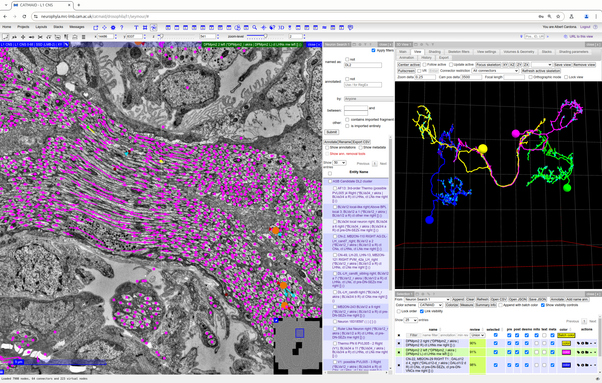
What can you do with a #CATMAID server? Say, let's look at the #Drosophila (vinegar fly, often referred to as fruit fly) larval central nervous system, generously hosted by the #VirtualFlyBrain https://l1em.catmaid.virtualflybrain.org/?pid=1&zp=108250&yp=82961.59999999999&xp=54210.799999999996&tool=tracingtool&sid0=1&s0=2.4999999999999996&help=true&layout=h(XY,%20%7B%20type:%20%22neuron-search%22,%20id:%20%22neuron-search-1%22,%20options:%20%7B%22annotation-name%22:%20%22papers%22%7D%7D,%200.6) or the #Platynereis (a marine annelid) server from the Jekely lab https://catmaid.jekelylab.ex.ac.uk/

First, directly interact by point-and-click: open widgets, find neurons by name or annotations, fire up a graph widget and rearrange neurons to make a neat synaptic connectivity diagram, or an adjacency matrix, or look at neuron anatomy in 3D. Most text–names, numbers–are clickable and filterable in some way, such as regular expressions.

Second, interact from other software. Head to r-catmaid https://natverse.org/rcatmaid/ (part of the #natverse suite by Philipp Schlegel @uni_matrix, Alex Bates and others) for an R-based solution from the Jefferis lab at the #MRCLMB. Includes tools such as #NBLAST for anatomical comparisons of neurons (see paper by Marta Costa et al. 2016 https://www.sciencedirect.com/science/article/pii/S0896627316302653 ).

If R is not your favourite, then how about #python: the #navis package, again by the prolific @uni_matrix, makes it trivial, and works also within #Blender too for fancy 3D renderings and animations. An earlier, simpler version was #catpy by @csdashm https://github.com/ceesem/catpy , who also has examples on access from #matlab.

Third, directly from a #psql prompt. As in, why not? #SQL is quite a straightforward language. Of course, you'll need privileged access to the server, so this one is only for insiders. Similarly privileged is from an #ipython prompt initialized via #django from the command line, with the entire server-side API at your disposal for queries.

Fourth, and one of my favourites: from the #javascript console in the browser itself. There are a handful of examples here https://github.com/catmaid/CATMAID/wiki/Scripting but the possibilities are huge. Key utilities are the "fetchSkeletons" macro-like javascript function https://github.com/catmaid/CATMAID/wiki/Scripting#count-the-number-of-presynaptic-sites-and-the-number-of-presynaptic-connectors-on-an-axon and the NeuronNameService.getInstance().getName(<skeleton_id>) function.

Notice every #CATMAID server has its /apis/, e.g., at https://l1em.catmaid.virtualflybrain.org/apis/ will list all GET or REST server access points. Reach to them as you please. See the documentation: https://catmaid.readthedocs.io/en/stable/api.html

In short: the data is there for you to reach out to, interactively or programmatically, and any fine mixture of the two as you see fit.

CATMAID

Virtual Fly Brain

Welcome to Virtual Fly Brain (VFB) - an interactive tool for neurobiologists to explore the detailed neuroanatomy, neuron connectivity and gene expression of Drosophila melanogaster. Our goal is to make it easier for researchers to find relevant anatomical information and reagents. We integrate the neuroanatomical and expression data from the published literature, as well as image datasets onto the same brain template, making it possible to run cross searches, find similar neurons and compare image data on our 3D Viewer.

Virtual Fly Brain