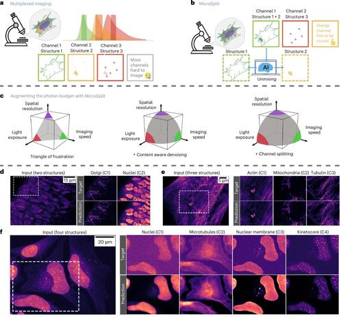
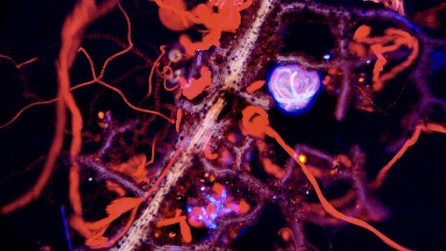
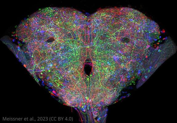
🔬 Explore types of microscopy 🧫—from simple to electron microscopes—unlocking cellular details for biology research and advanced diagnostics 🌱.
#Microscopy #TypesOfMicroscopy #BiologyLab #LifeScienceStudents #ElectronMicroscope #FluorescenceMicroscopy #CellBiology #Biotechnology #LaboratoryTechniques #BioResire
The aforementioned Synura. #fluorescencemicroscopy
Bluesky

Bluesky Social
Spring has sprung. Lots of euglena in Halls pond right now, making the red trails around the plant debris in the second pic (timelapse). So excited the ponds aren’t frozen any more. #sciart #microscopy #fluorescencemicroscopy
Bluesky

Bluesky Social
My install at Cornell’s department of natural resources, featuring a ‘rotifer altar.’ Gotta keep busy. #sciart #microscopy #fluorescencemicroscopy
Bluesky

Bluesky Social
Loving this fluorescent amoeba sped up 40x. I was on ‘amoeba time’ while on a zoom call with my former committee chair watching this fantastic creature slowly make its way around. #SciArt #microscopy #fluorescencemicroscopy #plankton
#Webinar Filamentous Bacteria: Causes, Impacts, Detection and Control. Insights into the biology and causes of filamentous bacteria, as well as their direct detection, impacts, and control strategies. #microscopy #wastewater #bacteria #filamentousbacteria #fluorescencemicroscopy #epifluorescence #vermicon https://www.vermicon.com/wastewater/events/webinar-filamentous-bacteria
Webinar: Filamentous bacteria

Scientists created a searchable #FluorescenceMicroscopy image resource of #Drosophila GAL4 driver expression patterns with single neuron resolution. Check out some of the images below. #Neuroscience #12DaysOfeLife https://elifesciences.org/articles/80660?utm_source=mastodon&utm_medium=social&utm_campaign=organic_12Days
A searchable image resource of Drosophila GAL4 driver expression patterns with single neuron resolution

Single neuron images of Drosophila driver lines reveal neuron shape and are searchable, enabling comparisons to electron microscopy and prediction of intersectional neuron targeting strategies.

eLife
Major advance in super-resolution fluorescence microscopy

Scientists led by Nobel Laureate Stefan Hell at the Max Planck Institute for Medical Research in Heidelberg have developed a super-resolution microscope with a spatio-temporal precision of one nanometer per millisecond. An improved version of their recently introduced MINFLUX super-resolution microscopy allowed tiny movements of single proteins to be observed at an unprecedented level of detail: the stepping motion of the motor protein kinesin-1 as it walks along microtubules while consuming ATP. The work, published in Science, highlights the power of MINFLUX as a revolutionary new tool for observing nanometer-sized conformational changes in proteins.

Phys.org