🚨 Are you at #MPS2023 and curious about cutting-edge brain-on-chip #biointerfaces?
Come by and have a chat with us! No matter if brain #organoids, #spheroids or acute tissue #slices, we have so much to show! 🔬
More about @3Brain's technology:
We empower researchers to explore #intelligent biological networks by linking them to #information technology.
#blogging about #neuroscience #networks #brain #semiconductor-based biointerfaces
#organizing the Massimo Grattarola Award to help young researchers get their ideas funded
| Website | www.3brain.com |
| Scicomm blog | https://www.3brain.com/pin/explorer-series |
| https://ch.linkedin.com/company/3brain |
🚨 Are you at #MPS2023 and curious about cutting-edge brain-on-chip #biointerfaces?
Come by and have a chat with us! No matter if brain #organoids, #spheroids or acute tissue #slices, we have so much to show! 🔬
More about @3Brain's technology:
Next week: Come explore our groundbreaking 3D microchips at #ISSCR2023 in Boston, USA!
No matter if #iPSC, primary neurons, or brain #organoids, @3Brain's #biointerface technology is revolutionizing neuroscience and advancing #stem cell research.

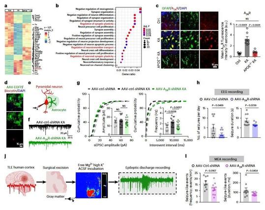

Chen et al. describe a new subtype of reactive astrocyte formed by APOE-mediated lipid accumulation in individuals with epilepsy and mouse models. These reactive astrocytes aggravate seizure symptoms and could serve as new therapeutic targets for epilepsy.
We are looking forward being at the next Organ-on-chip & organoids workshop in Zurich organized by @CSEMInfo !
@3Brain's Accura-3D chip was co-developed with the amazing talent at CSEM!
More info for the workshop:
https://www.csem.ch/en/events/next-gen-organ-on-chip-organoids-workshop/

The cerebellum is one of the most connected structures of the central nervous system and receives inputs over an extended frequency range. Nevertheless, the frequency dependence of cerebellar cortical processing remains elusive. In this work, we characterized cerebellar cortex responsiveness to mossy fibers activation at different frequencies and reconstructed the spread of activity in the sagittal and coronal planes of acute mouse cerebellar slices using a high-throughput high-density multielectrode array (HD-MEA). The enhanced spatiotemporal resolution of HD-MEA revealed the frequency dependence and spatial anisotropy of cerebellar activation. Mossy fiber inputs reached the Purkinje cell layer even at the lowest frequencies, but the efficiency of transmission increased at higher frequencies. These properties, which are likely to descend from the topographic organization of local inhibition, intrinsic electroresponsiveness, and short-term synaptic plasticity, are critical elements that have to be taken into consideration to define the computational properties of the cerebellar cortex and its pathological alterations.
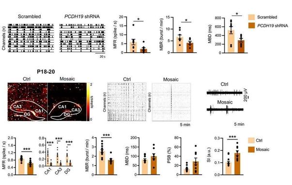

Mutations in PCDH19 gene, which encodes protocadherin-19 (PCDH19), cause Developmental and Epileptic Encephalopathy 9 (DEE9). Heterogeneous loss of PCDH19 expression in neurons is considered a key determinant of the disorder; however, how PCDH19 mosaic expression affects neuronal network activity and circuits is largely unclear. Here, we show that the hippocampus of Pcdh19 mosaic mice is characterized by structural and functional synaptic defects and by the presence of PCDH19-negative hyperexcitable neurons. Furthermore, global reduction of network firing rate and increased neuronal synchronization have been observed in different limbic system areas. Finally, network activity analysis in freely behaving mice revealed a decrease in excitatory/inhibitory ratio and functional hyperconnectivity within the limbic system of Pcdh19 mosaic mice. Altogether, these results indicate that altered PCDH19 expression profoundly affects circuit wiring and functioning, and provide new key to interpret DEE9 pathogenesis.
Exciting news!
@3Brain's @DenizPhd will soon come to Canada! We'll be showcasing our innovative high-density 3D #biointerface technology, a game-changer in neural complexity research.
Can't wait to connect at the 16th @CAN_ACN conference!
#3Brain #BrainTech #

Unraveling epilepsy mysteries w/ @3Brain's cutting-edge #biointerface tech! 🧠🌀
A study by Boucher-Routhier et al. ⬇️ used #GANs to analyze rare #spiral wave activity in cortical networks, key to understanding epilepsy.
Our high-res #microchip tech enabled detailed monitoring, paving the way for new treatment insights! 🚀🔬 #seizure #neuralcomplexity
Please read:
https://www.ncbi.nlm.nih.gov/pmc/articles/PMC10039524/

In the cerebral cortex, disinhibited activity is characterized by propagating waves that spread across neural tissue. In this pathological state, a widely reported form of activity are spiral waves that travel in a circular pattern around a fixed spatial ...
Interested in groundbreaking #biointerfaces for functional recording of #neuronal activity in 2D and #3D model systems? #organoids
Meet @denizphd at #FRM2023 ☀️ Algarve ☀️ !
Also: If you are a #neuroscience PhD/PostDoc currently looking for opportunities, feel free to come by!
How to rapidly screen for #neuromodulating compounds?
Free cultured cortical #spheroids 🧠 offer quick control over cell composition & functional testing
➡️ Coupled with @3Brain's #biointerface tech for drug screening & disease modeling 🔬
#stemcells

With the advent of human-induced pluripotent stem cells (hiPSCs) and differentiation protocols, methods to create in-vitro human-derived neuronal networks have been proposed. Although monolayer cultures represent a valid model, adding three-dimensionality (3D) would make them more representative of an in-vivo environment. Thus, human-derived 3D structures are becoming increasingly used for in-vitro disease modeling. Achieving control over the final cell composition and investigating the exhibited electrophysiological activity is still a challenge. Thence, methodologies to create 3D structures with controlled cellular density and composition and platforms capable of measuring and characterizing the functional aspects of these samples are needed. Here, we propose a method to rapidly generate neurospheroids of human origin with control over cell composition that can be used for functional investigations. We show a characterization of the electrophysiological activity exhibited by the neurospheroids by using micro-electrode arrays (MEAs) with different types (i.e., passive, C-MOS, and 3D) and number of electrodes. Neurospheroids grown in free culture and transferred on MEAs exhibited functional activity that can be chemically and electrically modulated. Our results indicate that this model holds great potential for an in-depth study of signal transmission to drug screening and disease modeling and offers a platform for in-vitro functional testing.
