Giving #minflux a whirl.
Easily setup, Google Reader integration allowing the Classic Reeder app to play nicely, no dramas.
(1/7) Proud to announce the preprint presenting my work on a fast, robust and scaleable polarisation interferometer for 3D #MINFLUX featuring dual-colour multi-emitter tracking in 3D!
https://www.biorxiv.org/content/10.1101/2023.12.09.570565v1
I am hiring !!
1 year Engineer position in optical microscopy to build a home – made RASTMIN microscope.

#superresolution #minflux #Microscopy
more info
https://integrativebiophysicsofmembranes.wordpress.com/2022/11/28/now-hiring/
Now hiring !

Several of our research projects have been recently funded by ANR & FranceBioImaging. Therefore we are now looking for two candidates, to fulfill the following positions : Postdoc position Post…

Team Integrative Biophysics of Membranes

Great explanation of #MINFLUX and how you can access this cutting-edge #bioimaging technique!

#ImagingScientist

@focalplane_jcs @the_node @LDN_PostdocNet @RMS_EarlyCareer @RoyalMicroSoc @Official_BSCB @_BSDB_ @BBSRC @The_MRC @STFC_Matters @UKRI_News @JofMicroscopy
---
RT @EuroBioImaging
Are you studying structural biology in cells🦠🧬 - or biological processes at the molecular level? Christopher Tynan, staff scientist at OCTOPUS, @C…
https://twitter.com/EuroBioImaging/status/1635954193667088385

Tweet / Twitter

Twitter

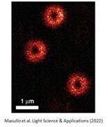
Chances are you don't have a #MINFLUX microscope and expert in your lab!

Don't let that stop you if you want to distinguish objects that are <10nm apart!

Apply to access MINFLUX at OCTOPUS, Harwell via @EuroBioImaging and get expert support for your project! #TooGoodToBeTrue
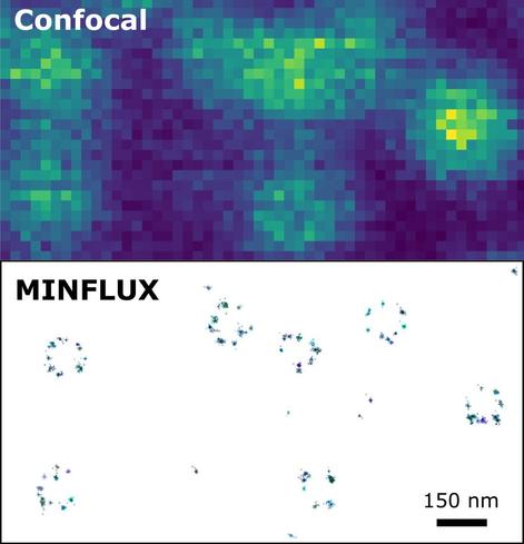
---
RT @EuroBioImaging
Want to see objects that are merely 5 nm apart - or track them in live cells with high temporal resolution? Give #MINFLUX a try🔬! Chr…
https://twitter.com/EuroBioImaging/status/1635642779542990848

Tweet / Twitter

Twitter
MPI pushed the #MINFLUX technique to the next level. It now allows protein dynamics to be observed under physiological conditions with a nanometer/millisecond.spatio-temporal precision of one
MINFLUX captures the rapid stepping motion of the motor protein kinesin-1 on a microtubule with nanometer precision.
@Albert
Ticker of #thenewuniverse, creator of https://fedi.science
Video: https://youtu.be/q6QAFacewf0
PR: https://www.mr.mpg.de/14582127/fast-even-at-the-nanometer-level
SCIENCE issue:
https://www.science.org/doi/10.1126/science.ade2650
Fediverse.science & Fedi.science

Fediverse.science unified with Fedi.science. The global fediverse of sciences, using both names alternatively suitable for each unser name

Mastodon hosted on fediverse.science

Tim Salditt (http://twitter.com/SaldittLab, substituting Jasper Frohn): #Multiscale #Xray Phase Contrast #Tomography at #GINIX/P10: Concepts, Implementation and Applications

- fantastic collaboration with the P10 team
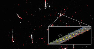
- directly comparing #STED to #Minflux; old #confocal? not worth mentioning 😂
- overview tomo, then zoom-in
- tumorous human pancreatic tissue #biopsy: quantify the #tumor type
- from electron density to metrics, quantify fibres etc.
- #hippocampus patho punch, #Alzheimer sample

Salditt Lab (@SaldittLab) / Twitter

We study soft matter & biomolecular assemblies, X-ray optics & holographic phase contrast X-ray imaging. Institute for X-Ray Physics (IRP) @uniGoettingen

Twitter
Dank einer bahnbrechenden Technik lassen sich im Fluoreszenzmikroskop lebende Strukturen auf den Nanometer genau auflösen und Proteinbewegungen im Millisekundentakt verfolgen.
Fluoreszenzmikroskopie: Die letzte Grenze
#Fluoreszenzmikroskopie #StefanHell #Superauflösung #HöchstauflendeMikroskope #Kinesin #Proteindynamik #Kernpore #STED-Mikroskopie #MINFLUX #MINSTED #Nobelpreis #Mikroskopie #Analytik #Physik #Biologie #Chemie #ITTech
Fluoreszenzmikroskopie: Die letzte Grenze

Mit speziellen Fluoreszenzmikroskopen lassen sich lebende Strukturen nanometergenau auflösen

2014 erhielt Stefan Hell den Nobelpreis für Chemie für die Entwicklung der STED-Mikroskopie. Jetzt hat er auf dieser Basis ein neues revolutionäres Mikroskopieverfahren ersonnen.
»Ein Potenzial, das noch in keiner Form gehoben ist«
#STED #Mikroskopie #MINFLUX #MINSTED #Fluoreszenzmikroskopue #PALMSTORM #Nobelpreis #Kinesin #Kinesin-1 #Nanoskopie #Hochauflösend #Kernpore #Kernporenkomplex #Physik #Biologie
»Ein Potenzial, das noch in keiner Form gehoben ist«

2014 erhielt Stefan Hell den Nobelpreis für Chemie für die Entwicklung der STED-Mikroskopie. Jetzt hat er auf dieser Basis ein neues revolutionäres Mikroskopieverfahren ersonnen.

Spektrum.de