24 Followers
30 Following
13 Posts
🌱🪴🌿🔬🏳️‍🌈
Lover of plants and microscopy. Research Scientist at Biology Department, Washington University in St. Louis. Part of the amazing Haswell lab. @radinbio on Twitter and Instagram.

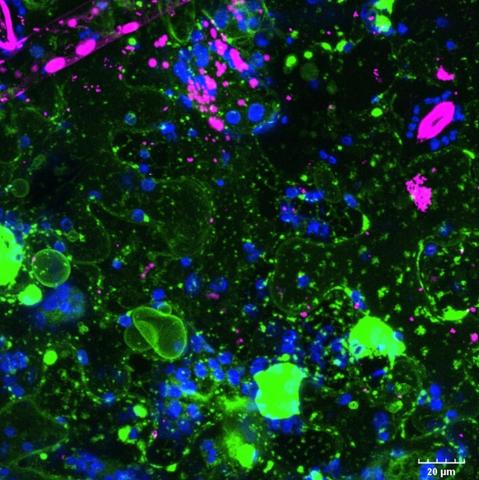
Not all microscopy images turn out great. Nevertheless, they can still be cool. This one definitely has an abstract art vibe. 🖼️
#Microscopy #fluoresence #cellbiology

The image shows Zmax projection of tobacco leaf, transiently overexpressing vacuolar membrane localized AtPIEZO1 tagged with EGFP. The EGFP fluorescence is in green. The leaf was also stained, unsuccessfully, with Mitotracker, in magenta. The chlorophyll autofluorescence is blue.

The images were made with the THUNDER Imager Model Organism dissecting scope (M205FCA) from @leicamicrosystems available at the WUSTL Biology department imaging core, using the 1x/0.03 objective with a 4x mag, greyscale camera, and below the sample illumination. The collage was made in Illustrator.

When moss cells are protoplasted, they behave like spores and can rebuild the whole plant. Over four days, protoplasts, when cultured on osmotically supported media, regenerate their cell walls and differentiate into chloronema cells, which slowly start to grow and divide.

When moved to regular media, the plants start to rapidly grow and divide. Within 3 days plants expand from 1-2 cells to what you see in the image.
At one point, some chloronema cells differentiate into caulonema cells.

Are these little aliens👽 or baby moss plants🪴? 😄
.
.
.
This collage is a great example of the power of plant regeneration. Each of these individual 7-day-old moss (Physcomitrium patens) plants, was regenerated from a single protoplast (plant cell with removed cell walls).

I love working with moss! They are even more adorable up close.
This is a single gametophore with the accompanying rhizoids from moss Physcomitrium patens. The image was made with the Olympus SZX7 dissecting scope with a DP71 camera from Olympus/Evident.

The image shows a single green gametophore from moss Physcomitrium patens. Gametophore is composed of many interlaced leaf-like structures, phyllids. Long and brownish rhizoids are protruding from the base of the gametophore.

This is what you see when you zoom in on fungus, growing uninvitedly on my "sterile" moss plates. What a beautiful hyphal network. #Fungi #Beatifulcontamination #Imaging
The image was made with the THUNDER Imager Model Organism dissecting scope (M205FCA) from @leicamicrosystems, available at the WUSTL Biology department imaging core. The image is a stitched composite from 9 (3 by 3) tile images captured using the 1x/0.6 objective with a 6.3x mag and greyscale camera.
Lab holiday party at the STL Art Museum. As I wind down my time as a PI, I’m more and more grateful for the folks I’ve been lucky to surround myself with. @radinbio and Josh are the best people and fantastic scientists!