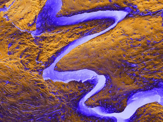
The bad bug hiding in the bush 🪲

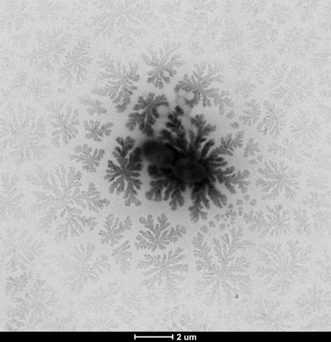
Antibiotic-resistant Acinetobacter baumannii hiding in a flower-like bed of crystallized buffer, exposed by the probing electrons of TEM.

👤 Dea Müller
 Bacterial Mechanobiology Laboratory, Persat Lab
 #TEM #BioTalos take at #CIME @EPFL
 https://www.epfl.ch/research/facilities/cime/index-html/image-gallery/image-of-the-moment/

#MicroscopyMonday #OpenScience #ElectronMicroscopy #CreativeCommons #MicroscopieÉlectronique #CCBYSA

🎉 Good news everyone !

The #CIME @EPFL electron microscopy image contest now requires participants to submit their work under a #CreativeCommons license. 🔬

 https://www.epfl.ch/research/facilities/cime/index-html/image-gallery/image-of-the-moment/

Result: These images can now circulate freely including here on the #fediverse!

So I’ll take the opportunity to share a few here. ✨

#ElectronMicroscopy #OpenScience #MicroscopyMonday #SEM #TEM

Image of the moment

The winner of March/April 2026 contest period is: “The bad bug hiding in the bush” by Dea Müller (Bacterial Mechanobiology Laboratory, Persat Lab). Congratulations! See all 2026 competitors gallery. Do you want to participate? Read contest rules and submit your image(s) here. See all 2025 competitors gallery. See all 2024 competitors gallery

EPFL

🎉 Bonne nouvelle, tout le monde !

Le concours d’images de microscopie électronique du #CIME @EPFL demande désormais que les participants soumettent leurs œuvres sous licence #CreativeCommons. 🔬

 https://www.epfl.ch/research/facilities/cime/index-html/image-gallery/image-of-the-moment/

Résultat : ces images pourront circuler librement, y compris ici sur la #fediverse !

Je vais donc en profiter pour en partager quelques-unes ici. ✨

#MicroscopieÉlectronique #ScienceOuverte #MicroscopyMonday #SEM #TEM

Image of the moment

The winner of March/April 2026 contest period is: “The bad bug hiding in the bush” by Dea Müller (Bacterial Mechanobiology Laboratory, Persat Lab). Congratulations! See all 2026 competitors gallery. Do you want to participate? Read contest rules and submit your image(s) here. See all 2025 competitors gallery. See all 2024 competitors gallery

EPFL

Whole-mouse imaging the hard way: Clearing, then slicing + block-face imaging with beam-scan / sensor scan-line synchronization and continuous stage movement in an oblique imaging setup (ViSOR).
This time, they scaled it up to whole-mouse and used pre-cleared mouse bodies (ARCHmap-blockface-ViSOR):

High-speed mapping of whole-mouse peripheral nerves at subcellular resolution
Shi et al., preprint at biorxiv 2025
https://doi.org/10.1101/2025.01.22.632569

#neuroscience #lightsheet #microscopy #MicroscopyMonday

Here's a #MicroscopyMonday segmentation of some neurons from the optic lobe, or visual processing area of the #Drosophila fruit fly's brain. The #connectome for the full optic lobe was released in 2024 by #HHMIJanelia, Google, and the University of Cambridge, with support from the Wellcome Trust.
This segmentation of #MicroscopyMonday data reconstructs neurons of the "MANC", or male adult nerve cord, from the #Drosophila fruit fly. The nerve cord is like a spinal cord and connects the brain and muscles. #HHMIJanelia, Google, and the University of Cambridge released this work in 2023.
A use of #MicroscopyMonday data is making segmentations. Here's one, of #Drosophila protocerebral bridge and nodulus neurons, from the 2020 Hemibrain by #HHMIJanelia and Google. Michał Januszewski and Viren Jain's team made an initial segmentation with #AI flood-fill algorithms...

Ya boi is back with more microscope photos :3

Here are a few pics of a cool looking filamentous algae I found looking at more bog water under the microscope. I don't know too much about algae species (yet!) so I can't say much in terms of identification for this specimen, but it's certainly pretty to look at!

#microscopy #microscopymonday #algae (does this really count as #NaturePhotography)

#MicroscopyMonday – No this is NOT an aerial view of the Grand Canyon.

This is a scanning electron microscopy image of the cranial sutures in the rat skull. 🔬💀🐀 These are fibrous joints where the bones of the skull interdigitate, allowing for flexibility and growth during infancy and early childhood before fusing in adulthood.

🔬🦴🧪

#bone #electronmicroscopy #imaging #biomaterials #science #medicine #biology #development #osteology #evolution

Happy #MicroscopyMonday!
Here is a kymograph of a cell crawling into a microchannel. Each horizontal row of pixels represents a line drawn from outside (green) through a channel (purple) at 5 second timepoints. ~1/2 way down (20 min) a cell fully enters the channel and takes off!