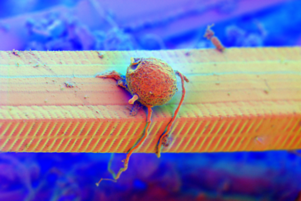
X-rays reveal kingfisher feather structure in unprecedented detail

Synchrotron radiation imaging revealed a porous, almost sponge-like nanostructure to create bright hues...

Ars Technica
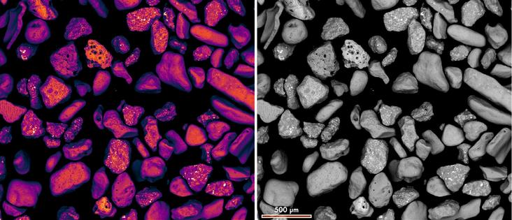
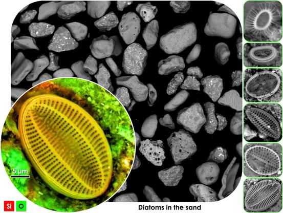
#ScanningElectronMicroscopy (SEM) and Energy Dispersive Spectroscopy (EDS) analysis help in high-resolution imaging and elemental composition analysis of materials. SEM Analysis Labs in Chennai offer precise microstructural evaluation for industries like electronics, metallurgy, and biomedical research, aiding in quality control and failure analysis.
https://mts-india.in/sem-eds/
SEM Analysis Lab In Chennai | SEM EDS Analysis

Scanning electron microscopy (SEM) analyzes the surface of materials in our SEM analysis lab in Chennai. SEM measures and evaluates surface pitting, failure analysis, characterization of dust, etc.

MTS India
πŸ”¬πŸ–οΈ Grains of #sand under a scanning electron #microscope – varied shapes, textures, vibrant microstructures. In the backscattered electron (BSE) #imaging mode, bright spots indicate presence of higher atomic number elements or higher average atomic density πŸ” #Microscopy #MicroscopicWorld #ElectronMicroscopy #ScanningElectronMicroscopy #Small

The other kind of imaging I do.

Not always in color but this one I like. And called Mike because of Monsters Inc

HFW = 32Β΅m

#ScanningElectronMicroscopy #SciArt
#ElectronMicroscopy
#SEM

Diatoms on #sand grains visualized using scanning electron microscopy! #Diatoms are a type of microalgae that have intricate and delicate silica shells. Scanning electron microscopy provides incredible detail, allowing us to appreciate the intricacies of #diatom morphology. #ScanningElectronMicroscopy #microscopy #naturephotography #scienceart #microscopicworld #microscopy #biology #lifesciences #silica #science #sciencemastodon #biomineral #biomaterials

– "Strange but beautiful" (@JimiHendrix)