192 Followers
53 Following
28 Posts
Dad. Microscopist. Photographer.
Director of OiVM Microscopy Core, Baylor College of Medicine http://youtube.com/@OiVM
Websitehttps://www.mileskirk.com/
OiVM Microscopy Corehttps://bcm.edu/oivm
Happy #MicroscopyMonday!
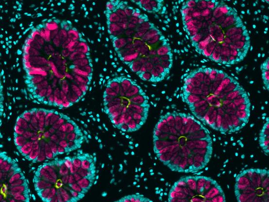
Here's Naegleria gruberi as a dividing amoeba and as a flagellate
Cellular space ship #FluorescenceFriday #microscopy
RT @ZebrafishRock
Migration of the lateral line sensory system during early zebrafish development. Credit to @damiandn. #ZebrafishZunday
Purkinje and lichen. Common shapes. #sciart
Happy belated Valentine's Day to everyone with this peculiar Ewing's sarcoma cell with a heart-shaped nucleus❤️ Magic mirror on the wall, who is the fairest one of all? DAPI = pink, Phalloidin = white

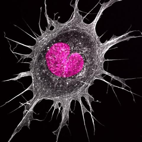
Destruction is the precursor to creation

Dividing cells disassembling their nuclear lamina during mitosis. One can also see the beautiful corrugated NE surrounded by ER
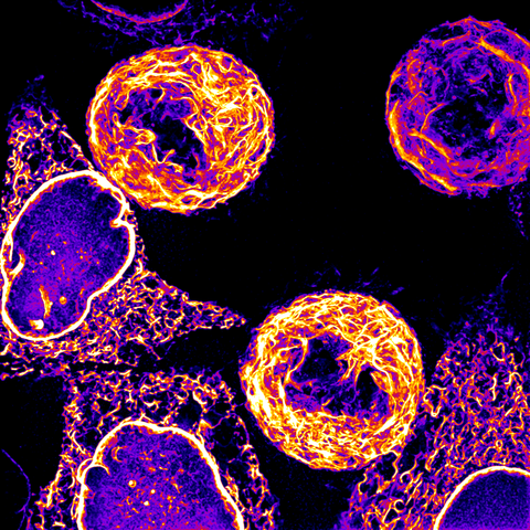
Taken during Zeiss demo organised by @PPhotonic @ZEISS_Group #Microscopy #FluorescenceFriday

This feels appropriate for the second thing I've ever posted to mastodon.

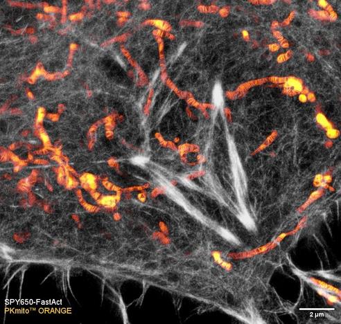
Have a good #FluorescenceFriday with this live HeLa cell imaged by #STED. Actin in white and mitochondria in "Duo Intense Yellow" LUT by James Manton.

🙏 Image Credit: Tianyan Liu

#PKmito #SPYprobes #imaging #lifesciences

Here is some eye candy of Merkel cells (green) and scales (magenta) to celebrate!