I am glad to say that the first manuscript from the lab is now available on BioRxiv
https://www.biorxiv.org/content/10.1101/2025.10.30.685496v1
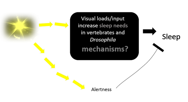
We set out to answer a simple question:
If the fruit fly #Drosophila cannot see clearly, how would they #sleep ?
1/8
For a naive 1st year Biology student, one would think such questions must have been answered long before, considering vision and light sensing are one of the most studied in Drosophila? But it was not the case around 2020s. Instead, opposing evidence showing *that vision can either suppress or enhance sleep depending on the experimental design:
https://doi.org/10.1093/sleep/zsz102
https://www.frontiersin.org/articles/10.3389/fphys.2020.00993/full
2/8

This is perhaps not surprising as vision are known to maintain one’s wakefulness and the same time providing sleep drive.

We therefore decided to look at the sleep profiles for all the classic mutant lines that affects phototransduction, plus those with defects in histamine transmission as histamine is the major neurotransmitter released by Drosophila photoreceptors.
3/8

To make sure the mutants we acquired retain their defects in phototransduction, the first author Yu-Chien Hung teamed up with Ben Warren at Keele University (https://warrenlab.wordpress.com/about-us/) to record electroretinogram.
4/8
Since we used a locomotion-based method to define sleep in Drosophila we also employed a long-forgotten analysis: waking activity, a simple ratio of locomotor activity over wake period, so we can be confident any increased amounts of sleep in mutants are not a result of loss of locomotion (eg http://www.jneurosci.org/content/29/35/11029.abstract)
5/8
The Effects of Caffeine on Sleep in Drosophila Require PKA Activity, But Not the Adenosine Receptor

Caffeine is one of the most widely consumed stimulants in the world and has been proposed to promote wakefulness by antagonizing function of the adenosine A2A receptor. Here, we show that chronic administration of caffeine reduces and fragments sleep in Drosophila and also lengthens circadian period. To identify the mechanisms underlying these effects of caffeine, we first generated mutants of the only known adenosine receptor in flies ( dAdoR ), which by sequence is most similar to the mammalian A2A receptor. Mutants lacking dAdoR have normal amounts of baseline sleep, as well as normal homeostatic responses to sleep deprivation. Surprisingly, these mutants respond normally to caffeine. On the other hand, the effects of caffeine on sleep and circadian rhythms are mimicked by a potent phosphodiesterase inhibitor, IBMX (3-isobutyl-1-methylxanthine). Using in vivo fluorescence resonance energy transfer imaging, we find that caffeine induces widespread increase in cAMP levels throughout the brain. Finally, the effects of caffeine on sleep are blocked in flies that have reduced neuronal PKA activity. We suggest that chronic administration of caffeine promotes wakefulness in Drosophila , at least in part, by inhibiting cAMP phosphodiesterase activity.

Journal of Neuroscience
So, what did we find?
1) Strikingly, seven of the eight tested mutants with mutations in four different phototransduction genes showed less sleep or shorter sleep bouts in the day.
2) artificially hyperpolarising eye photoreceptor also cause similar sleep loss in the day.
This finding supports that Drosophila phototransduction play a role in day sleep drive. We believe this small investigation provides a good reference point to untangle the role of sensory inputs in Drosophila sleep.
6/8

This work is a team effort from undergraduates, Erasmus, MSc, PhD and postdocs from the lab, thank you Yu-Chien, Mehran @mehranakhtar Xinghua @lixinghua Steven, Nithish, Clelia, Tobias and Jashmine for making this investigation possible!

#Neuroscience 7/8

also to add, the power of #openscience and PhD thesis deposition also lead us to realise others also find the same! so our results are reproducible* already :)
https://theses.hal.science/tel-04916427v1
Regulation of Sleep-wake rhythms by light in Drosophila melanogaster : Cellular and molecular histaminergic pathways from the visual System to clock neurons

Circadian docks orchestrate daily rhythms of physiological and behavioral processes in almost ail living organisms. This régulation relies on a molecular mechanism featuring transcriptional feedback loops, resulting in oscillations of clock gene expression. In Drosophi/a melanogaster, about 150 clock neurons form a self-sustaining mechanism, synchronizing through inter-neuronal communication to create a unified neuronal network and robust behavioral rhythms. A key aspect is the clock's ability to synchronize with day-night cycles by using external eues, or zeitgebers, with light being the most influential. Under 12/12 light-dark cycle, flies are crepuscular with morning and evening activity peaks during dawn and dusk, separated by daytime sleep aka siesta and nighttime sleep. Drosophila circadian photoreception involves two pathways: one utilizing the blue-light-sensitive photoreceptor Cryptochrome (Cry) expressed in approximately half of clock neurons, and the other involving the visual System expressing Rhodopsins.This thesis explores the régulation of the sleep-wake cycle and clock resetting through light input pathways, specifically focusing on the visual System. It includes three parts. The first part aimed at unravelling how Rhodopsin-dependent light pathways reset the circadian oscillator at the molecular level in the different clock neurons. Although it is known that light-activated Cry binds to the Timeless (Tim) clock protein, leading to circadian molecular clock resetting, the mechanism employed by visual System pathways remains elusive. Using cry mutants and a prolonged light puise protocol, the study analysed the initial molecular changes in clock neurons after light exposure. Results demonstrated that the visual System pathway also targets Tim, with its levels decreasing in response to light. The second part focused on the rôle of individual Rhodopsin-expressing photoreceptors in circadian entrainment under different light intensifies. Multi-mutant flies expressing only one of the six rhodopsins were used to discern their contributions. In low light conditions, where the NorpA-dependent phototransduction pathway is active, Rh1 and Rh6 emerged as the primary contributors to entrainment. Contrastingly, in high light conditions involving both canonical and non-canonical phototransduction pathways, ail six rhodopsin- expressing photoreceptors were found to provide entrainment, with Rh1, Rh5, and Rh6 being the most efficient in this process. The third part of this study focuses on the régulation of sleep via light in Drosophila, where sleep is characterized by sustained periods of quiescence, or 5-minute inactivity intervals associated with an increased arousal threshold. This régulation is orchestrated by the interplay of two processes: the homeostatic System and the circadian System. The research delves into the rôle of light, particularly through the light receptors Cry and the visual System, under high light intensity conditions. Additionally, the study explores the involvement of different phototransduction pathways and histaminergic downstream pathways in this sleep régulation process. The findings suggest that both Cry and the visual System are sufficient and required for the proper functioning of sleep. In contrast to entrainment, the canonical phototransduction pathway and the two histamine receptors are required to regulate the siesta effectively.

Also, we do have a #PhD position open to continue this investigation with the aid of #connectome in collaboration with Nils Reinhard @nils-reinhard.bsky.social
See details at https://www.kofanchenlab.net/come-join-us
Come Join US

Fully funded PhD positions available Application deadline: 27 November 2025 for MIBTP / 11 December for CSC

This manuscript is now published in
https://www.sciencedirect.com/science/article/pii/S2451994426000040
thanks again for everyone's hard work and the funding supports from #BBSRC and University of Leicester F50/F100 programme