https://www.scinexx.de/news/geowissen/gadolinium-kontrastmittel-verunreinigen-nord-und-ostsee/
A new study published in #EuropeanRadiology aimed to develop a #gadolinium-free #MRI-based diagnosis prediction tree for adult-type diffuse gliomas. (Aynur Azizova et al.)
Objectives To develop a gadolinium-free MRI-based diagnosis prediction decision tree (DPDT) for adult-type diffuse gliomas and to assess the added value of gadolinium-based contrast agent (GBCA) enhanced images. Materials and methods This study included preoperative grade 2–4 adult-type diffuse gliomas (World Health Organization 2021) scanned between 2010 and 2021. The DPDT, incorporating eleven GBCA-free MRI features, was developed using 18% of the dataset based on consensus readings. Diagnosis predictions involved grade (grade 2 vs. grade 3/4) and molecular status (isocitrate dehydrogenase (IDH) and 1p/19q). GBCA-free diagnosis was predicted using DPDT, while GBCA-enhanced diagnosis included post-contrast images. The accuracy of these predictions was assessed by three raters with varying experience levels in neuroradiology using the test dataset. Agreement analyses were applied to evaluate the prediction performance/reproducibility. Results The test dataset included 303 patients (age (SD): 56.7 (14.2) years, female/male: 114/189, low-grade/high-grade: 54/249, IDH-mutant/wildtype: 82/221, 1p/19q-codeleted/intact: 34/269). Per-rater GBCA-free predictions achieved ≥ 0.85 (95%-CI: 0.80–0.88) accuracy for grade and ≥ 0.75 (95%-CI: 0.70–0.80) for molecular status, while GBCA-enhanced predictions reached ≥ 0.87 (95%-CI: 0.82–0.90) and ≥ 0.77 (95%–CI: 0.71–0.81), respectively. No accuracy difference was observed between GBCA-free and GBCA-enhanced predictions. Group inter-rater agreement was moderate for GBCA-free (0.56 (95%-CI: 0.46–0.66)) and substantial for GBCA-enhanced grade prediction (0.68 (95%-CI: 0.58–0.78), p = 0.008), while substantial for both GBCA-free (0.75 (95%-CI: 0.69–0.80) and GBCA-enhanced (0.77 (95%-CI: 0.71–0.82), p = 0.51) molecular status predictions. Conclusion The proposed GBCA-free diagnosis prediction decision tree performed well, with GBCA-enhanced images adding little to the preoperative diagnostic accuracy of adult-type diffuse gliomas. Key Points Question Given health and environmental concerns, is there a gadolinium-free imaging protocol to preoperatively evaluate gliomas comparable to the gadolinium-enhanced standard practice? Findings The proposed gadolinium-free diagnosis prediction decision tree for adult-type diffuse gliomas performed well, and gadolinium-enhanced MRI demonstrated only limited improvement in diagnostic accuracy. Clinical relevance Even inexperienced raters effectively classified adult-type diffuse gliomas using the gadolinium-free diagnosis prediction decision tree, which, until further validation, can be used alongside gadolinium-enhanced images to respect standard practice, despite this study showing that gadolinium-enhanced images hardly improved diagnostic accuracy. Graphical Abstract
Educational Review: Review of strategies to reduce the contamination of the water #environment by #gadolinium-based contrast agents. (Helena M. Dekker et al.)
Gadolinium-based contrast agents (GBCA) are essential for diagnostic MRI examinations. GBCA are only used in small quantities on a per-patient basis; however, the acquisition of contrast-enhanced MRI examinations worldwide results in the use of many thousands of litres of GBCA per year. Data shows that these GBCA are present in sewage water, surface water, and drinking water in many regions of the world. Therefore, there is growing concern regarding the environmental impact of GBCA because of their ubiquitous presence in the aquatic environment. To address the problem of GBCA in the water system as a whole, collaboration is necessary between all stakeholders, including the producers of GBCA, medical professionals and importantly, the consumers of drinking water, i.e. the patients. This paper aims to make healthcare professionals aware of the opportunity to take the lead in making informed decisions about the use of GBCA and provides an overview of the different options for action.In this paper, we first provide a summary on the metabolism and clinical use of GBCA, then the environmental fate and observations of GBCA, followed by measures to reduce the use of GBCA. The environmental impact of GBCA can be reduced by (1) measures focusing on the application of GBCA by means of weight-based contrast volume reduction, GBCA with higher relaxivity per mmol of Gd, contrast-enhancing sequences, and post-processing; and (2) measures that reduce the waste of GBCA, including the use of bulk packaging and collecting residues of GBCA at the point of application.Critical relevance statement This review aims to make healthcare professionals aware of the environmental impact of GBCA and the opportunity for them to take the lead in making informed decisions about GBCA use and the different options to reduce its environmental burden.Key points• Gadolinium-based contrast agents are found in sources of drinking water and constitute an environmental risk.• Radiologists have a wide spectrum of options to reduce GBCA use without compromising diagnostic quality.• Radiology can become more sustainable by adopting such measures in clinical practice. Graphical Abstract
In this study published in #InsightsIntoImaging, Josef Vymazal and Aaron M. Rulseh conclude that #gadolinium-based contrast agents may accumulate in the brain; however, no adverse #neurological manifestations were detected relating to gadolinium retention.
Abstract Gadolinium-based contrast agents (GBCA) were introduced with high expectations for favorable efficacy, low nephrotoxicity, and minimal allergic-like reactions. Nephrogenic systemic fibrosis and proven gadolinium retention in the body including the brain has led to the restriction of linear GBCAs and a more prudent approach regarding GBCA indication and dosing. In this review, we present the chemical, physical, and clinical aspects of this topic and aim to provide an equanimous and comprehensive summary of contemporary knowledge with a perspective of the future. In the first part of the review, we present various elements and compounds that may serve as MRI contrast agents. Several GBCAs are further discussed with consideration of their relaxivity, chelate structure, and stability. Gadolinium retention in the brain is explored including correlation with the presence of metalloprotein ferritin in the same regions where visible hyperintensity on unenhanced T1-weighted imaging occurs. Proven interaction between ferritin and gadolinium released from GBCAs is introduced and discussed, as well as the interaction of other elements with ferritin; and manganese in patients with impaired liver function or calcium in Fahr disease. We further present the concept that only high-molecular-weight forms of gadolinium can likely visibly change signal intensity on unenhanced T1-weighted imaging. Clinical data are also presented with respect to potential neurological manifestations originating from the deep-brain nuclei. Finally, new contrast agents with relatively high relaxivity and stability are introduced. Critical relevance statement GBCA may accumulate in the brain, especially in ferritin-rich areas; however, no adverse neurological manifestations have been detected in relation to gadolinium retention. Key Points Gadolinium currently serves as the basis for MRI contrast agents used clinically. No adverse neurological manifestations have been detected in relation to gadolinium retention. Future contrast agents must advance chelate stability and relativity, facilitating lower doses. Graphical Abstract
This recent systematic review from #EuropeanRadiology highlighted the role of late #gadolinium enhancement (LGE) as the key #cardiac MR feature for arrhythmia risk stratification in patients with mitral valve prolapse (MVP). (Marco Gatti et al.)
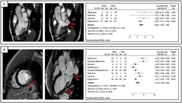
Objectives To perform a systematic review and meta-analysis of studies investigating the diagnostic value of cardiac magnetic resonance (CMR) features for arrhythmic risk stratification in mitral valve prolapse (MVP) patients. Materials and methods EMBASE, PubMed/MEDLINE, and CENTRAL were searched for studies reporting MVP patients who underwent CMR with assessment of: left ventricular (LV) size and function, mitral regurgitation (MR), prolapse distance, mitral annular disjunction (MAD), curling, late gadolinium enhancement (LGE), and T1 mapping, and reported the association with arrhythmia. The primary endpoint was complex ventricular arrhythmias (co-VAs) as defined by any non-sustained ventricular tachycardia, sustained ventricular tachycardia, ventricular fibrillation, or aborted sudden cardiac death. Meta-analysis was performed when at least three studies investigated a CMR feature. PROSPERO registration number: CRD42023374185. Results The meta-analysis included 11 studies with 1278 patients. MR severity, leaflet length/thickness, curling, MAD distance, and mapping techniques were not meta-analyzed as reported in < 3 studies. LV end-diastolic volume index, LV ejection fraction, and prolapse distance showed small non-significant effect sizes. LGE showed a strong and significant association with co-VA with a LogORs of 2.12 (95% confidence interval (CI): [1.00, 3.23]), for MAD the log odds-ratio was 0.95 (95% CI: [0.30, 1.60]). The predictive accuracy of LGE was substantial, with a hierarchical summary ROC AUC of 0.83 (95% CI: [0.69, 0.91]) and sensitivity and specificity rates of 0.70 (95% CI: [0.41, 0.89]) and 0.80 (95% CI: [0.67, 0.89]), respectively. Conclusions Our study highlights the role of LGE as the key CMR feature for arrhythmia risk stratification in MVP patients. MAD might complement arrhythmic risk stratification. Clinical relevance statement LGE is a key factor for arrhythmogenic risk in MVP patients, with additional contribution from MAD. Combining MRI findings with clinical characteristics is critical for evaluating and accurately stratifying arrhythmogenic risk in MVP patients. Key Points MVP affects 2–3% of the population, with some facing increased risk for arrhythmia. LGE can assess arrhythmia risk, and MAD may further stratify patients. CMR is critical for MVP arrhythmia risk stratification, making it essential in a comprehensive evaluation.
Mihály Károlyi et al. demonstrate that late #gadolinium enhancement (LGE) monitoring in #myocarditis is dependent on technique, finding that the full width at half maximum (FWHM) method yields the most consistent fibrotic tracking results.
Objectives To compare the repeatability and interrelation of various late gadolinium enhancement (LGE) assessment techniques for monitoring fibrotic changes in myocarditis follow-up. Materials and methods LGE extent change between baseline and 3-month cardiovascular magnetic resonance (CMR) was compared in patients with acute myocarditis using the full width at half maximum (FWHM), gray-scale thresholds at 5 and 6 standard deviations (SD5 and SD6), visual assessment with threshold (VAT) and full manual (FM) techniques. In addition, visual presence score (VPS), visual transmurality score (VTS), and a simplified visual change score (VCS) were assessed. Intraclass-correlation (ICC) was used to evaluate repeatability, and methods were compared using Spearman’s correlation. Results Forty-seven patients (38 male, median age: 27 [IQR: 21; 38] years) were included. LGE extent change differed among quantitative techniques (p < 0.01), with variability in the proportion of patients showing LGE change during follow-up (FWHM: 62%, SD5: 74%, SD6: 66%, VAT: 43%, FM: 60%, VPS: 53%, VTS: 77%, VCS: 89%). Repeatability was highest with FWHM (ICC: 0.97) and lowest with SD5 (ICC: 0.89). Semiquantitative scoring had slightly lower values (VPS ICC: 0.81; VTS ICC: 0.71). VCS repeatability was excellent (ICC: 0.93). VPS and VTS correlated with quantitative techniques, while VCS was positively associated with VPS, VTS, VAT, and FM, but not with FWHM, SD5, and SD6. Conclusion FWHM offers the least observer-dependent LGE follow-up after myocarditis. VPS, VTS, and VCS are practical alternatives, showing reliable correlations with quantitative methods. Classification of patients exhibiting either stable or changing LGE relies on the assessment technique. Clinical relevance statement This study shows that LGE monitoring in myocarditis is technique-dependent; the FWHM method yields the most consistent fibrotic tracking results, with scoring-based techniques as reliable alternatives. Key Points Recognition of fibrotic changes during myocarditis follow-up is significantly influenced by the choice of the quantification technique employed. The FWHM technique ensures highly repeatable tracking of myocarditis-related LGE changes. Segment-based visual scoring and the simplified visual change score offer practical, reproducible alternatives in resource-limited settings.
Review: Ten years of #gadolinium retention and deposition - ESMRMB-GREC looks backward and forward. (Aart J. van der Molen et al.)
Read the full review here 👉 https://link.springer.com/article/10.1007/s00330-023-10281-3
Abstract In 2014, for the first time, visible hyperintensities on unenhanced T1-weighted images in the nucleus dentatus and globus pallidus of the brain were associated with previous Gadolinium-based contrast agent (GBCA) injections and gadolinium deposition in patients with normal renal function. This led to a frenzy of retrospective studies with varying methodologies that the European Society of Magnetic Resonance in Medicine and Biology Gadolinium Research and Educational Committee (ESMRMB-GREC) summarised in 2019. Now, after 10 years, the members of the ESMRMB-GREC look backward and forward and review the current state of knowledge of gadolinium retention and deposition. Clinical relevance statement Gadolinium deposition is associated with the use of linear GBCA but no clinical symptoms have been associated with gadolinium deposition. Key Points • Traces of Gadolinium-based contrast agent-derived gadolinium can be retained in multiple organs for a prolonged time. • Gadolinium deposition is associated with the use of linear Gadolinium-based contrast agents. • No clinical symptoms have been associated with gadolinium deposition.