Insights Into Imaging

@InsightsImaging
31 Followers
2 Following
903 Posts
Insights into Imaging is a gold open access journal owned by the European Society of Radiology and edited by Editor-in-Chief, Prof. Luis MartΓ­-BonmatΓ­.

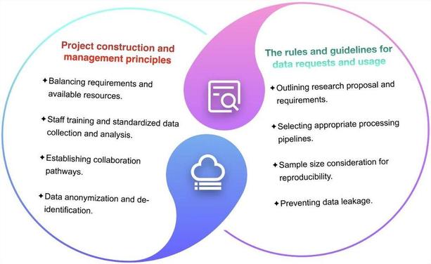
Xiang Fei et al. found that vascular morphology on high-frame rate contrast-enhanced ultrasound (H-CEUS) can indicate the risk of microvascular invasion (MVI), Ki-67 expression, and recurrence. This imaging technique offers insights into prognosis prediction prior to surgery.

#InsightsIntoImaging

πŸ”— https://buff.ly/4171yhO

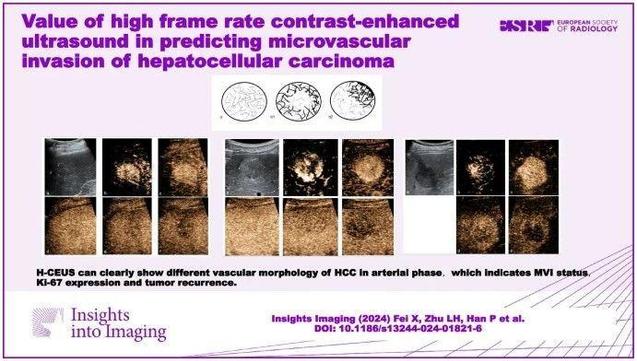
Value of high frame rate contrast-enhanced ultrasound in predicting microvascular invasion of hepatocellular carcinoma - Insights into Imaging

Objectives To investigate the value of vascular morphology on high frame rate contrast-enhanced ultrasound (H-CEUS) and CEUS Li-RADS in predicting microvascular invasion (MVI), Ki-67 expression and recurrence of hepatocellular carcinoma (HCC). Methods This retrospective study enrolled 78 patients with single HCC diagnosed by postoperative pathology between January 1, 2021, and June 30, 2022. All patients underwent ultrasound and H-CEUS examination before operation. H-CEUS image features and CEUS Li-RADS were compared in different MVI status and Ki-67 level. Multiple logistic regression analysis was performed to select independent variables for MVI. Differences in recurrence among different H-CEUS image features, MVI status and Ki-67 level were further analyzed. Results Tumor shape, vascular morphology, LR-M category, necrosis and AFP level were different between the MVI-positive group and MVI-negative group (p < 0.05). Vascular morphology and LR-M category were independent risk factors related to MVI (p < 0.05). Vascular morphology was also different between the high Ki-67 expression group and low Ki-67 expression group (p < 0.05). Vascular morphology, MVI status and Ki-67 expression were different between the recurrence group and no recurrence group (p < 0.05). Conclusion The vascular morphology of HCC on H-CEUS can indicate the risk of MVI status, Ki-67 expression and recurrence, which provides a feasible imaging technique for predicting the prognosis before operation. Critical relevance statement H-CEUS shows the different vascular morphology of HCC in arterial phase and indicates the risk of MVI, Ki-67 expression and recurrence, which provides a feasible imaging technique for clinician to judge the risk of MVI pre-operation and adopt appropriate treatment. Key Points H-CEUS can clearly show different vascular morphology of HCC in arterial phase. Vascular morphology on H-CEUS is associated with MVI status, Ki-67 expression and HCC recurrence. Preoperative MVI and Ki-67 expression prediction could help surgeons choose a more appropriate treatment plan. Graphical Abstract

SpringerOpen

Critical Review: Constructing and exploring neuroimaging projects - a survey from clinical practice to scientific research. (Ziyan Chen et al.)

#InsightsIntoImaging

πŸ”— https://buff.ly/417fqsA

Constructing and exploring neuroimaging projects: a survey from clinical practice to scientific research - Insights into Imaging

Abstract Over the past decades, numerous large-scale neuroimaging projects that involved the collection and release of multimodal data have been conducted globally. Distinguished initiatives such as the Human Connectome Project, UK Biobank, and Alzheimer’s Disease Neuroimaging Initiative, among others, stand as remarkable international collaborations that have significantly advanced our understanding of the brain. With the advancement of big data technology, changes in healthcare models, and continuous development in biomedical research, various types of large-scale projects are being established and promoted worldwide. For project leaders, there is a need to refer to common principles in project construction and management. Users must also adhere strictly to rules and guidelines, ensuring data safety and privacy protection. Organizations must maintain data integrity, protect individual privacy, and foster stakeholders’ trust. Regular updates to legislation and policies are necessary to keep pace with evolving technologies and emerging data-related challenges. Critical relevance statement By reviewing global large-scale neuroimaging projects, we have summarized the standards and norms for establishing and utilizing their data, and provided suggestions and opinions on some ethical issues, aiming to promote higher-quality neuroimaging data development. Key Points Global neuroimaging projects are increasingly advancing but still face challenges. Constructing and utilizing neuroimaging projects should follow set rules and guidelines. Effective data management and governance should be developed to support neuroimaging projects. Graphical Abstract

SpringerOpen

This retrospective study assessed the diagnostic performance of prostate MRI by estimating the proportion of clinically significant #ProstateCancer (csPCa) in patients without prostate pathology. It found varying csPCa proportions based on PI-RADS scores, with sensitivity of 76.6–77.3%, specificity of 67.5–78.6%, and NPV of 84.4–87.2%. (Hirotsugu Nakai et al.)

#InsightsIntoImaging #PIRADS

πŸ”— https://buff.ly/4fHwrOm

Estimated diagnostic performance of prostate MRI performed with clinical suspicion of prostate cancer - Insights into Imaging

Purpose To assess the diagnostic performance of prostate MRI by estimating the proportion of clinically significant prostate cancer (csPCa) in patients without prostate pathology. Materials and methods This three-center retrospective study included prostate MRI examinations performed for clinical suspicion of csPCa (Grade group β‰₯ 2) between 2018 and 2022. Examinations were divided into two groups: pathological diagnosis within 1 year after the MRI (post-MRI pathology) is present and absent. Risk prediction models were developed using the extracted eleven common predictive variables from the patients with post-MRI pathology. Then, the csPCa proportion in the patients without post-MRI pathology was estimated by applying the model. The area under the receiver operating characteristic curve (AUC), sensitivity, specificity, and positive and negative predictive values (PPV/NPV) of prostate MRI in diagnosing csPCa were subsequently calculated for patients with and without post-MRI prostate pathology (estimated statistics) with a positive threshold of PI-RADS β‰₯ 3. Results Of 12,191 examinations enrolled (mean age, 65.7 years Β± 8.4 [standard deviation]), PI-RADS 1–2 was most frequently assigned (55.4%) with the lowest pathological confirmation rate of 14.0–18.2%. Post-MRI prostate pathology was found in 5670 (46.5%) examinations. The estimated csPCa proportions across facilities were 12.6–15.3%, 18.4–31.4%, 45.7–69.9%, and 75.4–88.3% in PI-RADS scores of 1–2, 3, 4, and 5, respectively. The estimated (observed) performance statistics were as follows: AUC, 0.78–0.81 (0.76–0.79); sensitivity, 76.6–77.3%; specificity, 67.5–78.6%; PPV, 49.8–66.6% (52.0–67.7%); and NPV, 84.4–87.2% (82.4–86.6%). Conclusion We proposed a method to estimate the probabilities harboring csPCa for patients who underwent prostate MRI examinations, which allows us to understand the PI-RADS diagnostic performance with several metrics. Clinical relevance statement The reported estimated performance metrics are expected to aid in understanding the true diagnostic value of PI-RADS in the entire prostate MRI population performed with clinical suspicion of prostate cancer. Key Points Calculating performance metrics only from patients who underwent prostate biopsy may be biased due to biopsy selection criteria, especially in PI-RADS 1–2. The estimated area under the receiver operating characteristic curve of PI-RADS in the entire prostate MRI population ranged from 0.78 to 0.81 at three facilities. The estimated statistics are expected to help us understand the true PI-RADS performance and serve as a reference for future studies. Graphical Abstract

SpringerOpen

πŸ•―οΈ Join us over the next four weeks as we celebrate #rAdvent!

πŸ•―οΈ Each week starting December 1st, we'll take a deep dive into an article from the #ESRJournals family chosen by our Social Media Editorial Team, providing expert insight into some of our most popular articles published in our three journals!

Stay tuned...

#EuropeanRadiology
#InsightsIntoImaging
#EuropeanRadiologyExperimental

Educational Review: Roles of #MRI evaluation of pelvic recurrence in patients with #RectalCancer. (Patricia Perola Dantas et al.)

#InsightsIntoImaging

πŸ”— https://buff.ly/3ZkH2cw

Roles of MRI evaluation of pelvic recurrence in patients with rectal cancer - Insights into Imaging

Abstract Developments in the multidisciplinary treatment of rectal cancer with advances in preoperative magnetic resonance imaging (MRI), surgical techniques, neoadjuvant chemoradiotherapy, and adjuvant chemotherapy have had a significant impact on patient outcomes, increasing the rates of curative surgeries and reducing pelvic recurrence. Patients with pelvic recurrence have worse prognoses, with an impact on morbidity and mortality. Although local recurrence is more frequent within 2 years of surgical resection of the primary tumor, late recurrence may occur. Clinical manifestations can vary from asymptomatic, nonspecific symptoms, to pelvic pain, bleeding, and fistulas. Synchronous metastatic disease occurs in approximately 50% of patients diagnosed with local recurrence. MRI plays a crucial role in posttreatment follow-up, whether by identifying viable neoplastic tissues or acting as a tool for therapeutic planning and assessing the resectability of these lesions. Locally recurrent tissues usually have a higher signal intensity than muscle on T2-weighted imaging. Thus, attention is required for focal heterogeneous lesions, marked contrast enhancement, early invasive behavior, and asymmetric appearance, which are suspicious for local recurrence. However, postsurgical inflammatory changes related to radiotherapy and fibrosis make it difficult to detect initial lesions. This study therefore aimed to review the main imaging patterns of pelvic recurrence and their implications for the surgical decision-making process. Critical relevance statement MRI plays a crucial role in the posttreatment follow-up of rectal cancer, whether by identifying viable neoplastic tissues or by acting as a tool for therapeutic planning. This study reviewed the main imaging patterns of pelvic recurrence. Key Points MRI aids in surgical planning and the detection of pelvic recurrence and postoperative complications. Being familiar with surgical techniques enables radiologists to identify expected MRI findings. Patterns of rectal cancer recurrence have been categorized by pelvic compartments. Neoplastic tissue may mimic postsurgical and postradiotherapy changes. Resectability of pelvic recurrence is highly related to lesion location. Graphical Abstract

SpringerOpen

Nan Meng et al. show that glucose chemical exchange saturation transfer (glucoCEST) MRI, in combination with amide proton transfer-weighted imaging (APTWI) and diffusion-weighted imaging (DWI), can effectively differentiate between high- and low-grade #RectalCancer, which could be a promising imaging marker.

#InsightsIntoImaging

πŸ”— https://buff.ly/3Z6Jy4O

Glucose chemical exchange saturation transfer MRI for predicting the histological grade of rectal cancer: a comparative study with amide proton transfer-weighted and diffusion-weighted imaging - Insights into Imaging

Background To evaluate the utility of glucose chemical exchange saturation transfer (glucoCEST) MRI with non-contrast injection in predicting the histological grade of rectal cancer. Methods This prospective analysis included 60 patients with preoperative rectal cancer who underwent pelvic glucoCEST, amide proton transfer-weighted imaging (APTWI), and diffusion-weighted imaging (DWI). In total, 21 low-grade and 39 high-grade cases were confirmed by postoperative pathology. The MTRasym (1.2 ppm), MTRasym (3.5 ppm), and apparent diffusion coefficient (ADC) values of lesions between the low-grade and high-grade groups were compared. The area under the receiver operating characteristic curve (AUC) was generated to evaluate the diagnostic performance of each technique. Logistic regression (LR) analysis was applied to determine independent predictors and for multi-parameter combined diagnosis. Results Elevated MTRasym (1.2 ppm), MTRasym (3.5 ppm) values and lower ADC values were observed in the high-grade group compared with low-grade cases (all p < 0.01). The AUCs of MTRasym (1.2 ppm), MTRasym (3.5 ppm), and ADC for differentiating between low- and high-grade rectal cancer cases were 0.792, 0.839, and 0.855, respectively. The diagnostic performance of the combination of the three indexes was improved (AUC, 0.969; sensitivity, 95.24%; specificity, 87.18%). The good consistency and reliability of the combination of independent predictors were demonstrated by calibration curve analysis and DCA. Conclusion The glucoCEST MRI without contrast injection, APTWI, and DWI all facilitate the assessment of histological grade in rectal cancer, and the combination of the three can effectively discriminate between high- and low-grade rectal cancer, which is expected to be a promising imaging marker. Critical relevance statement The glucose chemical exchange saturation transfer MRI method facilitates the assessment of histological grade in rectal cancer and offers additional information to improve the diagnostic performance of amide proton transfer-weighted imaging, and diffusion-weighted imaging. Key Points Glucose chemical exchange saturation transfer imaging could differentiate histological grade. Amide proton transfer-weighted and diffusion-weighted were associated with histological grade. The combination of different parameters showed the best diagnostic performance. Graphical Abstract

SpringerOpen

This article in our #RadiologyAndBeyond series looks at a nationwide #Dutch survey which aimed to evaluate the diagnostic accuracy of #MRI, CT, and [18F]FDG-PET-CT in assessing lymph node involvement in clinically early-stage #CervicalCancer. (Ester P. Olthof et al.)

#InsightsIntoImaging #RadiologyAndBeyond

πŸ”— https://buff.ly/49w5BoS

Diagnostic accuracy of MRI, CT, and [18F]FDG-PET-CT in detecting lymph node metastases in clinically early-stage cervical cancer β€” a nationwide Dutch cohort study - Insights into Imaging

Objectives Imaging is increasingly used to assess lymph node involvement in clinically early-stage cervical cancer. This retrospective study aimed to evaluate the diagnostic accuracy of MRI, CT, and [18F]FDG-PET-CT. Methods Women with International Federation of Gynaecology and Obstetrics (FIGO) 2009 stage IA2-IIA cervical cancer and pretreatment imaging between 2009 and 2017 were selected from the Netherlands Cancer Registry. Patient-based and region-based (i.e. pelvic and common iliac) nodal status was extracted from radiology reports. Pathology results were considered the reference standard for calculating accuracy indices. Multiple imputation was used for missing pathology to limit verification bias risk. Results Nodal assessment was performed in 1676 patients with MRI, 926 with CT, and 379 with [18F]FDG-PET-CT, with suspicious nodes detected in 17%, 16%, and 48%, respectively. [18F]FDG-PET-CT was used to confirm MRI/CT results in 95% of patients. Pathology results were imputed for 30% of patients. [18F]FDG-PET-CT outperformed MRI and CT in detecting patient-based nodal metastases with sensitivities of 80%, 48%, and 40%, and AUCs of 0.814, 0.706, and 0.667, respectively, but not in specificity: 79%, 92%, and 92%. Region-based analyses showed similar indices in the pelvic region, but worse performance in the common iliac region with AUCs of 0.575, 0.554, and 0.517, respectively. Conclusions [18F]FDG-PET-CT outperformed MRI and CT in detecting nodal metastases, which may be related to its use as a verification modality. However, MRI and CT had the highest specificity. As MRI is generally performed routinely to assess local and regional spread of cervical cancer, [18F]FDG-PET-CT can be used to confirm suspicious nodes. Critical relevance statement Accurate assessment of the nodal status in clinically early-stage cervical cancer is essential for tumour staging, treatment decision making and prognosis. Key points β€’ The accuracy of MRI, CT or [18F]FDG-PET-CT for nodal staging in early cervical cancer is a subject of discussion. β€’ Overall, [18F]FDG-PET-CT outperformed MRI, followed by CT, when used as a verification modality. β€’ Staging with MRI and the addition of [18F]FDG-PET-CT to verify high-risk cases seems to be a good approach. Graphical Abstract

SpringerOpen

A recent #InsightsIntoImaging study assessing the #RIRADS system for evaluating #radiology requests in an #Italian cohort found most requests were inadequate, especially for routine imaging. RI-RADS demonstrated substantial reliability, supporting its use in clinical settings. (Marco Parillo et al.)

πŸ”— https://buff.ly/4i27Rtk

Assessment of Reason for Exam Imaging Reporting and Data System (RI-RADS) in inpatient diagnostic imaging referrals - Insights into Imaging

Objectives To test the Reason for Exam Imaging Reporting and Data System (RI-RADS) in assessing the quality of radiology requests in an Italian cohort of inpatients; to evaluate the interobserver reliability of RI-RADS. Methods A single-center quality care study was designed to retrospectively identify consecutive radiology request forms for computed tomography, magnetic resonance imaging, and conventional radiography examinations. One radiologist scored the requests using the RI-RADS. The association between RI-RADS and clinical request variables (urgent request, on-call requests, indication for imaging, requesting specialty, imaging modality, and body region) was evaluated. We calculated interobserver agreement between four readers in a subset of 450 requests. Results We included 762 imaging requests. RI-RADS grades A (adequate request), B (barely adequate request), C (considerably limited request), D (deficient request), and X were assigned to 8 (1%), 49 (7%), 237 (31%), 404 (53%), and 64 (8%) of cases, respectively. In the multivariate analysis, the indication for imaging, body region, and requesting specialty significantly influenced the RI-RADS. Indications for imaging with a high risk of poor RI-RADS grade were routine preoperative imaging and device check requests. The upper extremity was the body region with the highest risk of poor RI-RADS grade. Requesting specialties with a high risk of poor RI-RADS grade were cardiovascular surgery, intensive care medicine, and orthopedics. The analysis of the interobserver agreement revealed substantial agreement for the RI-RADS grade. Conclusion The majority of radiology exam requests were inadequate according to RI-RADS, especially those for routine imaging. RI-RADS demonstrated substantial reliability, suggesting that it can be satisfactorily employed in clinical settings. Critical relevant statement The implementation of RI-RADS can provide a framework for standardizing radiology requests, thereby enabling quality assurance and promoting a culture of quality improvement. Key Points RI-RADS aims to grade the completeness of radiology requests. Over half of the imaging requests were RI-RADS D grade; RI-RADS demonstrated substantial reliability. Most radiology requests were inadequate and RI-RADS could classify them in clinical practice. Graphical Abstract

SpringerOpen

Last week, the "Eternal City" played host to the ESR's Annual Leadership Meeting, where our Editors-in-Chief discussed the state of their respective journals alongside Marc Dewey, Chair of the ESR Publications Committee, and Ioana Gheonea, Chair of the Young ESR Committee.

#EuropeanRadiology
#InsightsIntoImaging
#EuropeanRadiologyExperimental
#ESRJournals

Educational Review: Proceedings from an international consensus meeting on ablation in urogenital diseases. (Roberto Iezzi et al.)

#InsightsIntoImaging

πŸ”— https://buff.ly/3V1SSWk

Proceedings from an international consensus meeting on ablation in urogenital diseases - Insights into Imaging

Abstract Percutaneous image-guided ablation techniques are a consolidated therapeutic alternative for patients with high preoperative surgical risk for the management of oncological diseases in multiple body districts. Each technique has both pros and cons according to the type of energy delivered, mechanism of action, and site of application. The present article reviews the most recent literature results on ablation techniques applied in the field of genitourinary diseases (kidney, adrenal glands, prostate, and uterus), describing the advantages of the use of each technique and their technical limitations and summarizing the major recommendations from an international consensus meeting. Critical relevant statement The article critically evaluates the efficacy and safety of ablation therapies for various genitourinary tract diseases, demonstrating their potential to improve patient outcomes and advance clinical radiology by offering minimally invasive, effective alternatives to traditional surgical treatments. Key Points Ablation therapies are effective alternatives to surgery for renal cell carcinoma. Ablation techniques offer effective treatment for intermediate-risk prostate cancer. Ablation is a promising tool for adrenal tumor management. Ablation reduces fibroid symptoms and volume, offering an alternative to surgery. Graphical Abstract

SpringerOpen