Human Fovea Detector
https://www.shadertoy.com/view/4dsXzM
#HackerNews #Human #Fovea #Detector #Technology #Innovation #Vision #Science
Human Fovea Detector
https://www.shadertoy.com/view/4dsXzM
#HackerNews #Human #Fovea #Detector #Technology #Innovation #Vision #Science
5/We identified 912 bipolar cells in he HFseg1 volume, including the major types recognized in non-human primates, though with some notable differences (#connectomic, #retina, #fovea).
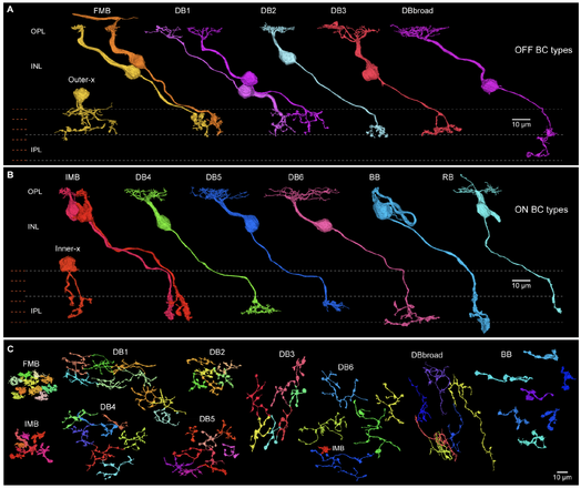

With over 10[14][1] synapses, the human brain presents a seemingly insurmountable challenge to a nano-scale circuit-level understanding of its diverse neural systems. The foveal retina however presents a feasible site for a complete connectome of a key human CNS structure. Foveal cells and circuits are miniaturized and compressed to densely sample the visual image at highest resolution to initiate form, color and motion perception. Here we use computational methods first applied to the fly brain to provide a draft connectome of all neurons in a foveal volume. We found synaptic connections distinct to humans linking short-wavelength sensitive cones to color vision pathways. Moreover, by reconstructing excitatory synaptic pathways arising from cone photoreceptors we found that 96% of foveal ganglion cells contribute to only three major pathways to the brain. This new resource reveals unique features of a human neural system and opens a door to its complete connectome. In Brief Deep-learning based reconstruction of cells and synapses in a human fovea reveals visual pathway origins and human-specific wiring for color vision, opening the door to a complete synaptic-level connectome of this critical locus in the human central nervous system. Highlights ### Competing Interest Statement K.L., N.K., D.I., T.N., R.L., S.P., A.H., J.A.B., J.S. and T.M. declare financial interests in Zetta AI. S.G. is owner of Aware LLC and developed the NeuroMaps.app. The remaining authors declare no competing interests. [1]: #ref-14
it often feels odd, like the sketched version is reality
Not odd at all! /You are experiencing how #perception works./
What we perceive is a #reality built up in parts from data the #fovea of our eye gathers flickering around and our brain mixing it with past experience and an a presumed understanding of our context (the model of our surroundings). This is why we can't find our keys when they are right in front of us: we expect them elsewhere and movement or a flash of metal didn't add the detail we needed to mentally construct the scene. We cannot see a reality we cannot mentally visualize!
What is happening is through your practice of seeing and rendering your #art is that your brain is constructing a sense of the art being real, or becoming real as you would while #painting or #drawing it. You are seeing the world as you want to #sketch it.
This is a good thing.
I have the same sense in my #photography. Two things I took away from my college drawing classes was being able to see negative spaces, shadows and volume, and being able to see white as colored by reflection or illumination, that is white as blue outside in the shade. When I look around, I see resultant photographs and know what will render and what won't, and when to lift the #lens.
I trust that sense, and I'm sure you do or will soon.
#BoostingIsSharing
#CommentingIsCool
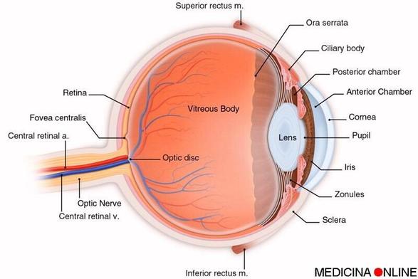
🔵 L’occhio quando fissa è fermo? Cosa sono i movimenti saccadici?
👉 Leggi l’articolo: https://medicinaonline.co/2017/02/12/locchio-quando-fissa-e-fermo-cosa-sono-i-movimenti-saccadici/
✅ #occhio #saccade #saccadici #sguardo #vista #fovea #retina #macula #EmilioAlessioLoiacono #MedicinaOnLine