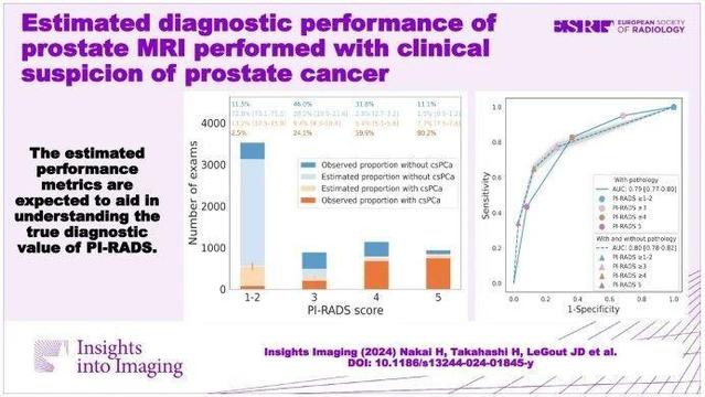
This retrospective study assessed the diagnostic performance of prostate MRI by estimating the proportion of clinically significant #ProstateCancer (csPCa) in patients without prostate pathology. It found varying csPCa proportions based on PI-RADS scores, with sensitivity of 76.6–77.3%, specificity of 67.5–78.6%, and NPV of 84.4–87.2%. (Hirotsugu Nakai et al.)

#InsightsIntoImaging #PIRADS

🔗 https://buff.ly/4fHwrOm

Estimated diagnostic performance of prostate MRI performed with clinical suspicion of prostate cancer - Insights into Imaging

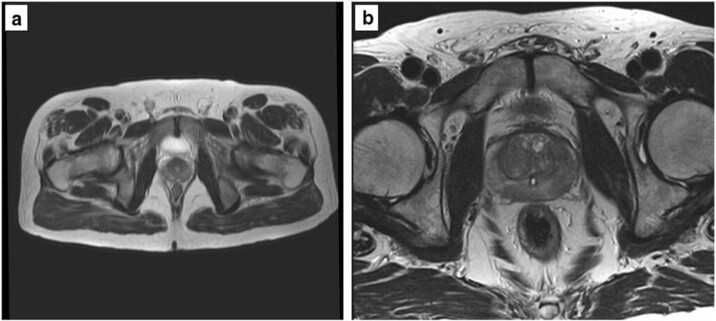
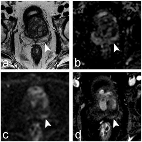
Purpose To assess the diagnostic performance of prostate MRI by estimating the proportion of clinically significant prostate cancer (csPCa) in patients without prostate pathology. Materials and methods This three-center retrospective study included prostate MRI examinations performed for clinical suspicion of csPCa (Grade group ≥ 2) between 2018 and 2022. Examinations were divided into two groups: pathological diagnosis within 1 year after the MRI (post-MRI pathology) is present and absent. Risk prediction models were developed using the extracted eleven common predictive variables from the patients with post-MRI pathology. Then, the csPCa proportion in the patients without post-MRI pathology was estimated by applying the model. The area under the receiver operating characteristic curve (AUC), sensitivity, specificity, and positive and negative predictive values (PPV/NPV) of prostate MRI in diagnosing csPCa were subsequently calculated for patients with and without post-MRI prostate pathology (estimated statistics) with a positive threshold of PI-RADS ≥ 3. Results Of 12,191 examinations enrolled (mean age, 65.7 years ± 8.4 [standard deviation]), PI-RADS 1–2 was most frequently assigned (55.4%) with the lowest pathological confirmation rate of 14.0–18.2%. Post-MRI prostate pathology was found in 5670 (46.5%) examinations. The estimated csPCa proportions across facilities were 12.6–15.3%, 18.4–31.4%, 45.7–69.9%, and 75.4–88.3% in PI-RADS scores of 1–2, 3, 4, and 5, respectively. The estimated (observed) performance statistics were as follows: AUC, 0.78–0.81 (0.76–0.79); sensitivity, 76.6–77.3%; specificity, 67.5–78.6%; PPV, 49.8–66.6% (52.0–67.7%); and NPV, 84.4–87.2% (82.4–86.6%). Conclusion We proposed a method to estimate the probabilities harboring csPCa for patients who underwent prostate MRI examinations, which allows us to understand the PI-RADS diagnostic performance with several metrics. Clinical relevance statement The reported estimated performance metrics are expected to aid in understanding the true diagnostic value of PI-RADS in the entire prostate MRI population performed with clinical suspicion of prostate cancer. Key Points Calculating performance metrics only from patients who underwent prostate biopsy may be biased due to biopsy selection criteria, especially in PI-RADS 1–2. The estimated area under the receiver operating characteristic curve of PI-RADS in the entire prostate MRI population ranged from 0.78 to 0.81 at three facilities. The estimated statistics are expected to help us understand the true PI-RADS performance and serve as a reference for future studies. Graphical Abstract

SpringerOpen

This study investigated if a #DeepLearning software could improve #PIRADS scoring consistency on bi-parametric prostate MRI. (Aydan Arslan et al.)

#InsightsIntoImaging

🔗 https://buff.ly/4c4hNP6

Does deep learning software improve the consistency and performance of radiologists with various levels of experience in assessing bi-parametric prostate MRI? - Insights into Imaging

Objective To investigate whether commercially available deep learning (DL) software improves the Prostate Imaging-Reporting and Data System (PI-RADS) scoring consistency on bi-parametric MRI among radiologists with various levels of experience; to assess whether the DL software improves the performance of the radiologists in identifying clinically significant prostate cancer (csPCa). Methods We retrospectively enrolled consecutive men who underwent bi-parametric prostate MRI at a 3 T scanner due to suspicion of PCa. Four radiologists with 2, 3, 5, and > 20 years of experience evaluated the bi-parametric prostate MRI scans with and without the DL software. Whole-mount pathology or MRI/ultrasound fusion-guided biopsy was the reference. The area under the receiver operating curve (AUROC) was calculated for each radiologist with and without the DL software and compared using De Long’s test. In addition, the inter-rater agreement was investigated using kappa statistics. Results In all, 153 men with a mean age of 63.59 ± 7.56 years (range 53–80) were enrolled in the study. In the study sample, 45 men (29.80%) had clinically significant PCa. During the reading with the DL software, the radiologists changed their initial scores in 1/153 (0.65%), 2/153 (1.3%), 0/153 (0%), and 3/153 (1.9%) of the patients, yielding no significant increase in the AUROC (p > 0.05). Fleiss’ kappa scores among the radiologists were 0.39 and 0.40 with and without the DL software (p = 0.56). Conclusions The commercially available DL software does not increase the consistency of the bi-parametric PI-RADS scoring or csPCa detection performance of radiologists with varying levels of experience.

SpringerOpen

Byung Kwan Park et al. explore a new transperineal ultrasound (TPUS)-guided biopsy technique that may contribute to the detection of large #PIRADS 5 #ProstateCancer in men following a Miles' operation.

#InsightsIntoImaging

🔗 https://buff.ly/4ebTNeC

New transperineal ultrasound-guided biopsy for men in whom PSA is increasing after Miles’ operation - Insights into Imaging

Objectives Currently, a prostate biopsy is guided by transrectal ultrasound (US) alone. However, this biopsy cannot be performed in men without an anus. The aim of this study was to show the outcomes of a new transperineal US (TPUS)-guided biopsy technique in patients who underwent Miles’ operation. Methods Between April 2009 and March 2022, TPUS-guided biopsy was consecutively conducted in 9 patients (median, 71 years; range, 61–78 years) with high prostate-specific antigen values (22.60 ng/mL; 6.19–69.7 ng/mL). Their anuses were all removed due to rectal cancer. TPUS-guided biopsy was performed according to information on prostate magnetic resonance imaging. The technical success rate, cancer detection rate, and complication rate were recorded. Tumor sizes were compared between benign and cancer groups using an unpaired t-test with Welch’s correction. Results The new TPUS-guided biopsy was successfully performed in all patients. Cancer was detected in 77.8% (7/9) of the patients. These were all categorized as PI-RADS 5. Among them, the detection rate of significant cancer (Gleason score 7 or higher) was 66.7% (6/9). The median tumor size was 2.4 cm (1.7–3.1 cm). However, two patients were diagnosed with benign tissue with PI-RADS 3 or PI-RADS 4. Their median tumor size was 1.0 cm (0.8–1.2 cm). There was significant difference between the cancer and benign groups (p = 0.037) in terms of tumor size. Neither post-biopsy bleeding nor infections occurred. Conclusions New TPUS-guided biopsy technique may contribute to detecting large PI-RADS 5 prostate cancer in men after Miles’ operation.

SpringerOpen

Zhoujie Sun et al. explore the diagnostic performance of targeted biopsy combined with regional systematic biopsy in patients with different #PIRADS and histologic zones for prostate lesions, which shows promise as an alternative approach.

#InsightsIntoImaging

🔗 https://buff.ly/3WSuAQk

Diagnostic performance of regional systematic biopsy for prostate cancer stratified by PI-RADS and histologic zones - Insights into Imaging

Objectives To explore the diagnostic performance of targeted biopsy (TB) combined with regional systematic biopsy (RSB) in patients with different Prostate Imaging Reporting and Data System (PI-RADS) and histologic zones for prostate lesions. Methods This retrospective study included 1301 patients who underwent multiparametric MRI followed by combined MRI/US fusion-guided TB+systematic biopsy (SB) between January 2019 and October 2022. RSB was defined as the four perilesional SB cores adjacent to an MRI-positive lesion. Cancer detection rates were calculated for TB + SB, TB, SB, and TB + RSB, while the McNemar test was utilized for multiple comparisons among them. Subgroup analyses were performed based on different Pl-RADS and histologic zones. Results Of 1301 included participants (median age, 68 years; interquartile range, 63–74 years), 16,104 total biopsy cores were performed. TB + RSB detected clinically significant prostate cancer in 70.9% (922/1301) of patients, which was significantly higher than TB (67.4%, p < 0.001) or SB (67.5%, p < 0.001) but similar to TB + SB (71.0%, p = 0.50). Compared with TB + SB, TB + RSB required fewer median biopsy cores (6.3 vs. 12.4, p < 0.001) and had a higher proportion of positive cores (56.3% vs. 39.0%, p < 0.001). Subgroup analysis showed that TB had outstanding sensitivity for detecting PI-RADS 5 lesions in the PZ. Conclusions Compared with TB + SB, TB + RSB achieved a similar clinically significant prostate cancer detection rate while requiring fewer biopsy cores and exhibiting higher diagnostic efficiency. Critical relevance statement For MRI-positive prostate lesions, targeted biopsy combined with regional systematic biopsy could serve as an alternative diagnostic approach to targeted biopsy combined with systematic biopsy. Key Points The scheme of prostate biopsy needs to be optimized. Regional systematic biopsy decreases the total number of cores taken. Targeted biopsies combined with regional systematic biopsies improve prostate diagnostic efficiency. Graphical Abstract

SpringerOpen

Quantitative #ultrasound shear wave elastography (USWE) and mpMRI using #PIRADS classification shows a good degree of prediction for Gleason score of clinically significant #ProstateCancer (csPCa). (Wael Ageeli et al.)

#InsightsIntoImaging #MRI

🔗 https://insightsimaging.springeropen.com/articles/10.1186/s13244-021-01039-w

Quantitative ultrasound shear wave elastography (USWE)-measured tissue stiffness correlates with PIRADS scoring of MRI and Gleason score on whole-mount histopathology of prostate cancer: implications for ultrasound image-guided targeting approach - Insights into Imaging

Objective To correlate quantitative tissue stiffness measurements obtained by transrectal ultrasound shear wave elastography (USWE) with PI-RADS scoring of multiparametric magnetic imaging resonance (mpMRI) using Gleason scores of radical prostatectomy as a reference standard. Patients and methods 196 men with localised prostate cancer were prospectively recruited into the study and had quantitative prostate tissue stiffness measurements in kilopascals (kPa) using transrectal USWE prior to radical prostatectomy. PI-RADS scores of mpMRI were also obtained in all the men. Imaging and histopathology of radical prostatectomy specimen were oriented to each other using patient specific customised 3D moulds to guide histopathology grossing of radical prostatectomy specimens. All included patients had confirmed PCa on TRUS-guided biopsies, had both USWE and mpMRI imaging data, and underwent radical prostatectomy. Chi-square test with 95% confidence interval was used to assess the difference between Gleason score (GS) of radical prostatectomy and PI-RADS classification, as well as GS of radical prostatectomy and stiffness (in Kpa) using USWE. The correlation coefficient (r) was calculated in order to investigate relation between PI-RADS classification and tissue stiffness in kPa. Results There was a statistically significant correlation between USWE-measured tissue stiffness and GS (χ2 (2, N = 196) = 23.577, p < 0.001). Also, there was a statistically significant correlation between Gleason score and PI-RADS score (χ2 (2, N = 196) = 12.838, p = 0.002). High PIRADS on MRI and high stiffness on USWE (> 100 kPa) detected more than 80% and 90% high risk prostate cancer disease. However, a weak correlation coefficient of 0.231 was observed between PI-RADS score and level of tissue stiffness measured in kPa. Conclusion Quantitative USWE and mpMRI using PI-RADS classification provide a good degree of prediction for Gleason score of clinically significant prostate cancer (csPCa). Stiffer lesions on ultrasound showed a weak correlation with PI-RADS scoring system. USWE could be used to target suspected prostate cancer.

SpringerOpen

Educational Review: Understanding PI-QUAL for #prostate #MRI quality: a practical primer for #radiologists. (Francesco Giganti et al.)

#InsightsIntoImaging #ProstateCancer #PIRADS

🔗 https://insightsimaging.springeropen.com/articles/10.1186/s13244-021-00996-6

Understanding PI-QUAL for prostate MRI quality: a practical primer for radiologists - Insights into Imaging

Prostate magnetic resonance imaging (MRI) of high diagnostic quality is a key determinant for either detection or exclusion of prostate cancer. Adequate high spatial resolution on T2-weighted imaging, good diffusion-weighted imaging and dynamic contrast-enhanced sequences of high signal-to-noise ratio are the prerequisite for a high-quality MRI study of the prostate. The Prostate Imaging Quality (PI-QUAL) score was created to assess the diagnostic quality of a scan against a set of objective criteria as per Prostate Imaging-Reporting and Data System recommendations, together with criteria obtained from the image. The PI-QUAL score is a 1-to-5 scale where a score of 1 indicates that all MR sequences (T2-weighted imaging, diffusion-weighted imaging and dynamic contrast-enhanced sequences) are below the minimum standard of diagnostic quality, a score of 3 means that the scan is of sufficient diagnostic quality, and a score of 5 implies that all three sequences are of optimal diagnostic quality. The purpose of this educational review is to provide a practical guide to assess the quality of prostate MRI using PI-QUAL and to familiarise the radiologist and all those involved in prostate MRI with this scoring system. A variety of images are also presented to demonstrate the difference between suboptimal and good prostate MR scans.

SpringerOpen

MULTI study: PI-RADSv2.1 descriptors are more specific than PI-RADSv2 descriptors when assessing the same set of #MRI lesions, but did not improve inter-reader variability. (Florian Di Franco et al.)

#InsightsIntoImaging #PIRADS #ProstateCancer

🔗 https://insightsimaging.springeropen.com/articles/10.1186/s13244-023-01391-z

Characterization of high-grade prostate cancer at multiparametric MRI: assessment of PI-RADS version 2.1 and version 2 descriptors across 21 readers with varying experience (MULTI study) - Insights into Imaging

Objective To assess PI-RADSv2.1 and PI-RADSv2 descriptors across readers with varying experience. Methods Twenty-one radiologists (7 experienced (≥ 5 years) seniors, 7 less experienced seniors and 7 juniors) assessed 240 ‘predefined’ lesions from 159 pre-biopsy multiparametric prostate MRIs. They specified their location (peripheral, transition or central zone) and size, and scored them using PI-RADSv2.1 and PI-RADSv2 descriptors. They also described and scored ‘additional’ lesions if needed. Per-lesion analysis assessed the ‘predefined’ lesions, using targeted biopsy as reference; per-lobe analysis included ‘predefined’ and ‘additional’ lesions, using combined systematic and targeted biopsy as reference. Areas under the curve (AUCs) quantified the performance in diagnosing clinically significant cancer (csPCa; ISUP ≥ 2 cancer). Kappa coefficients (κ) or concordance correlation coefficients (CCC) assessed inter-reader agreement. Results At per-lesion analysis, inter-reader agreement on location and size was moderate-to-good (κ = 0.60–0.73) and excellent (CCC ≥ 0.80), respectively. Agreement on PI-RADSv2.1 scoring was moderate (κ = 0.43–0.47) for seniors and fair (κ = 0.39) for juniors. Using PI-RADSv2.1, juniors obtained a significantly lower AUC (0.74; 95% confidence interval [95%CI]: 0.70–0.79) than experienced seniors (0.80; 95%CI 0.76–0.84; p = 0.008) but not than less experienced seniors (0.74; 95%CI 0.70–0.78; p = 0.75). As compared to PI-RADSv2, PI-RADSv2.1 downgraded 17 lesions/reader (interquartile range [IQR]: 6–29), of which 2 (IQR: 1–3) were csPCa; it upgraded 4 lesions/reader (IQR: 2–7), of which 1 (IQR: 0–2) was csPCa. Per-lobe analysis, which included 60 (IQR: 25–73) ‘additional’ lesions/reader, yielded similar results. Conclusions Experience significantly impacted lesion characterization using PI-RADSv2.1 descriptors. As compared to PI-RADSv2, PI-RADSv2.1 tended to downgrade non-csPCa lesions, but this effect was small and variable across readers.

SpringerOpen