We are sadly leaving Mastodon... but don't worry you can still find us on Bluesky (
https://bsky.app/profile/genomiqueens.bsky.social) and LinkedIn (
https://linkedin.com/company/genomique-ens/)!
(We have got some exciting updates coming so go check it out 👀👀)

GenomiqueENS (@genomiqueens.bsky.social)
Genomics core facility of IBENS @ibensens.bsky.social - Ecole Normale Supérieure @normalesup.bsky.social - @psl-univ.bsky.social
#Nanopore #transcriptomics #SingleCellRNAseq #eucaryotes #low-input
website: https://genomique.biologie.ens.fr/
New insights into chronic myelomonocytic leukemia: Combining short & long-read RNA sequencing with k-mer analysis unveils 4 chimeric RNAs. These results could help advance patient risk stratification & treatment! We collaborated with Institute for Regenerative Medicine and Biotherapy and the Centre National de Recherche en Génomique Humaine by constructing the libraries and performing @nanopore sequencing from human samples.
New study reshapes our understanding of P-bodies as active participants in RNA regulation, not just storage! We collaborated with the laboratoire de Biologie du Développement from @Sorbonne Université by constructing the libraries and performing
#illumina sequencing from human cell lines 🧬 🔬
https://doi.org/10.1101/2024.04.16.589748Excited to announce that our QC tool for
#Nanopore sequencers
#ToulligQC is now available as an EPI2ME-compatible workflow! 🎉🎉
The EPI2ME Desktop application is easy to use and allows users of any skill level to run their bioinformatic analyses!
Go check it out on our github
https://lnkd.in/eQPQbmST and EPI2ME Labs
https://lnkd.in/e26Bq44ALinkedIn
This link will take you to a page that’s not on LinkedIn
How do tardigrades survive ionizing radiation? With our collaborators from AVIV at MNHN, we uncovered DNA repair mechanisms in tardigrades by combining
#illumina and
#nanopore sequencing on 3 species of tardigrade.
https://doi.org/10.7554/eLife.92621.3
Comparative transcriptomics reveal a novel tardigrade-specific DNA-binding protein induced in response to ionizing radiation
When exposed to high-dose ionizing radiation, tardigrades undergo extensive DNA damage, like humans, but cope by stimulating expression of DNA repair proteins and of TDR1, a novel tardigrade-specific DNA-binding protein.
New transcriptomic study demonstrates Decitabine as potential antiviral compound against equine rhinopneumonitis! With our collaborators BioTargen, we performed
#illumina sequencing and RNA-seq analysis from horse samples.
https://doi.org/10.3390/v16050746

A Screening Study Identified Decitabine as an Inhibitor of Equid Herpesvirus 4 That Enhances the Innate Antiviral Response
Equid herpesvirus 4 (EHV-4) is a common respiratory pathogen in horses. It sporadically induces abortion or neonatal death. Although its contribution in neurological disorders is not clearly demonstrated, there is a strong suspicion of its involvement. Despite preventive treatments using vaccines against EHV-1/EHV-4, the resurgence of alpha-EHV infection still constitutes an important threat to the horse industry. Yet very few studies have been conducted on the search for antiviral molecules against EHV-4. A screening of 42 antiviral compounds was performed in vitro on equine fibroblast cells infected with the EHV-4 405/76 reference strain (VR2230). The formation of cytopathic effects was monitored by real-time cell analysis (RTCA), and the viral load was quantified by quantitative PCR. Aciclovir, the most widely used antiviral against alpha-herpesviruses in vivo, does not appear to be effective against EHV-4 in vitro. Potential antiviral activities were confirmed for eight molecules (idoxuridine, vidarabine, pritelivir, cidofovir, valganciclovir, ganciclovir, aphidicolin, and decitabine). Decitabine demonstrates the highest efficacy against EHV-4 in vitro. Transcriptomic analysis revealed the up-regulation of various genes implicated in interferon (IFN) response, suggesting that decitabine triggers the immune antiviral pathway.
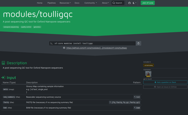
The report contains exhaustive information about the sequencing run, basecalling and demultiplexing steps => read count and length distributions, homogeneity of the run, location of potential flow cell spatial biases, statistics about pass and fail reads, data by barcodes ...
ToulligQC 2.7 => is fast (few minutes on a laptop) => supports all versions of Guppy and Dorado => can be used with all the @nanopore
sequencing devices hashtag#MinION hashtag#GridION hashtag#PrometION => accepts POD5, Fast5, FASTQ, BAM etc..
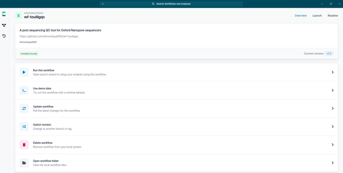
Pleased to announce our QC tool for
@nanopore sequencers
#ToulligQC is available as a
@nf_core module 🎉
Implemented in Nextflow, users can modulate ToulligQC to their need!
Exciting new research reveals fasting's impact on the blood-brain barrier! With our collaborators from Université Paris Cité Médicine, we performed
#illumina sequencing and RNA-seq analysis from samples of rats brain cells.
https://doi.org/10.1186/s12987-024-00526-8

Fasting upregulates the monocarboxylate transporter MCT1 at the rat blood-brain barrier through PPAR δ activation - Fluids and Barriers of the CNS
Background The blood-brain barrier (BBB) is pivotal for the maintenance of brain homeostasis and it strictly regulates the cerebral transport of a wide range of endogenous compounds and drugs. While fasting is increasingly recognized as a potential therapeutic intervention in neurology and psychiatry, its impact upon the BBB has not been studied. This study was designed to assess the global impact of fasting upon the repertoire of BBB transporters. Methods We used a combination of in vivo and in vitro experiments to assess the response of the brain endothelium in male rats that were fed ad libitum or fasted for one to three days. Brain endothelial cells were acutely purified and transcriptionaly profiled using RNA-Seq. Isolated brain microvessels were used to assess the protein expression of selected BBB transporters through western blot. The molecular mechanisms involved in the adaptation to fasting were investigated in primary cultured rat brain endothelial cells. MCT1 activity was probed by in situ brain perfusion. Results Fasting did not change the expression of the main drug efflux ATP-binding cassette transporters or P-glycoprotein activity at the BBB but modulated a restrictive set of solute carrier transporters. These included the ketone bodies transporter MCT1, which is pivotal for the brain adaptation to fasting. Our findings in vivo suggested that PPAR δ, a major lipid sensor, was selectively activated in brain endothelial cells in response to fasting. This was confirmed in vitro where pharmacological agonists and free fatty acids selectively activated PPAR δ, resulting in the upregulation of MCT1 expression. Moreover, dosing rats with a specific PPAR δ antagonist blocked the upregulation of MCT1 expression and activity induced by fasting. Conclusions Altogether, our study shows that fasting affects a selected set of BBB transporters which does not include the main drug efflux transporters. Moreover, we describe a previously unknown selective adaptive response of the brain vasculature to fasting which involves PPAR δ and is responsible for the up-regulation of MCT1 expression and activity. Our study opens new perspectives for the metabolic manipulation of the BBB in the healthy or diseased brain.