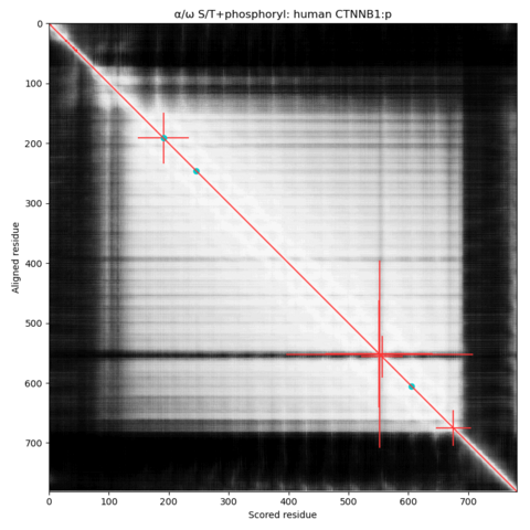
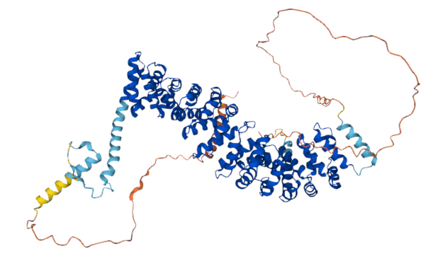
α⧸ω S/T phosphorylation diagram of human β-catenin (CTNNB1:p). The diagram is dominated by a large bright patch corresponding to a domain of armadillo repeats. The repeats are composed of anti-parallel α-helixes with rather short unstructured loops. The 3 S/T+phosphoryl acceptors are situated on these loops: the helixes themselves are (as usual) kinase-resistant.
#teamProteome
A similar diagram for ubiquitination acceptors shows that ubiquitin ligases are much less fussy about where they tag this molecule. Neither PTM seems to be interested in the IDRs on either end of the sequence.
The lack of PTMs on the C-terminal IDR may be caused by a long tryptic peptide (718-781) that is rarely observed.
The phospho-IDRs associated with helical repeat domains are restricted to the short loops connecting helixes, making the patterns resemble the classical phosphorylation "site" still illustrated in ye olde text books.