"Critical point drying of brain tissue for X-ray phase-contrast imaging", Khan et al. 2026 (Schaefer's and Bosch's labs).
https://journals.iucr.org/s/issues/2026/03/00/wuz5001/index.html

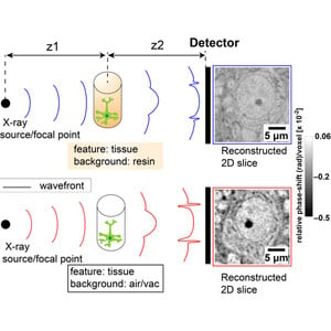
"we propose to replace interstitial material by air to enhance X-ray phase contrast of the ultrastructural features. Critical point drying (CPD) of heavy-metal-stained mouse brain tissue produced samples with preserved ultrastructure, a nanofoam-like material that remains compatible with follow-up conventional resin embedding. Using two synchrotron-based setups [...] we found that CPD samples consistently showed 2–4× stronger phase-shift signal than samples embedded in resin."

#neuroscience #XNH #synchrotron

Critical point drying of brain tissue for X-ray phase-contrast imaging

X-ray phase contrast tomography can efficiently image brain tissue at subcellular resolution. Critical point drying allows a gentle replacement of interstitial material by air, enhancing X-ray phase contrast of the ultrastructural features.

Journal of Synchrotron Radiation

"Functional mapping of the molluscan brain guided by synchrotron X-ray tomography", Crossley et al. 2025
https://pubmed.ncbi.nlm.nih.gov/40014565/

#neuroscience #Lymnaea #molluscs #XNH

Functional mapping of the molluscan brain guided by synchrotron X-ray tomography - PubMed

Molluscan brains are composed of morphologically consistent and functionally interrogable neurons, offering rich opportunities for understanding how neural circuits drive behavior. Nonetheless, detailed component-level CNS maps are often lacking, total neuron numbers are unknown, and organizational …

PubMed

Now that's a big deal, and from a very credible source:

"Self-supervised image restoration in coherent X-ray neuronal microscopy", Laugros et al. (Alexandra Pacureanu) 2025
https://www.biorxiv.org/content/10.1101/2025.02.10.633538v1.full

"we present a self-supervised image restoration approach that simultaneously improves spatial resolution, contrast, and data acquisition speed. This enables revealing synapses with XNH, marking a major milestone in the quest for generating connectomes of full mammalian brains."

X-ray nanoholography took a turn towards higher resolution and higher throughput.

#XNH #connectomics #neuroscience

Self-supervised image restoration in coherent X-ray neuronal microscopy

Coherent X-ray microscopy is emerging as a transformative technology for neuronal imaging, with the potential to offer a scalable solution for reconstruction of neural circuits in millimeter sized tissue volumes. Specifically, X-ray holographic nanoto-mography (XNH) brings together outstanding capabilities in terms of contrast, spatial resolution and data acquisition speed. While recent XNH developments already enabled generating valuable datasets for neuro-sciences, a major challenge for reconstruction of neural circuits remained overcoming resolving power limits to distinguish smaller neurites and synapses in the reconstructed volumes. Here we present a self-supervised image restoration approach that simultaneously improves spatial resolution, contrast, and data acquisition speed. This enables revealing synapses with XNH, marking a major milestone in the quest for generating connectomes of full mammalian brains. We demonstrate that this method is effective for various types of neuronal tissues and acquisition schemes. We propose a scalable implementation compatible with multi-terabyte image volumes. Altogether, this work brings large-scale X-ray nanotomography to a new precision level. ### Competing Interest Statement The authors have declared no competing interest.

bioRxiv