51st #NikonSmallWorld winners reveal a hidden world of wonders: https://zorz.it/PqBOh
#JeremyGray #51stAnnualNikonSmallWorldPhotomicrographyCompetition #macro #winners #micro #microscopy #Nikon #PhotoContest #PhotoMicrography
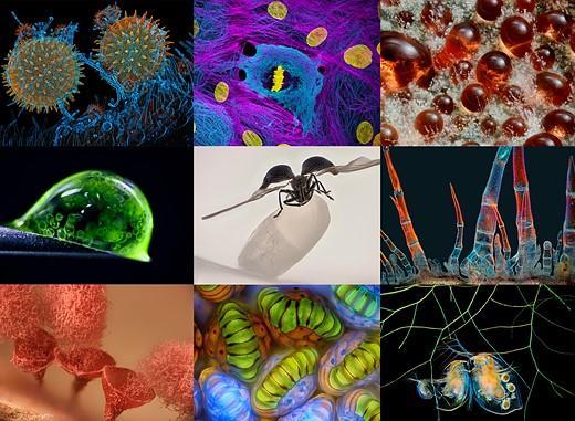
51st #NikonSmallWorld winners reveal a hidden world of wonders: https://zorz.it/PqBOh
#JeremyGray #51stAnnualNikonSmallWorldPhotomicrographyCompetition #macro #winners #micro #microscopy #Nikon #PhotoContest #PhotoMicrography
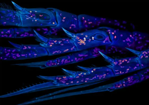
Marine copepod are small aquatic crustaceans.
Photograph: Zachary Sanchez/Nikon Small World
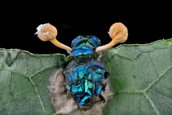
Parasitic fungus (Cordycipitaceae) on a fly (Calliphoridae). Cordycipitaceae can infect a Calliphoridae into a zombie-like state, leaving them to die and spread to other insects.
Photograph: Eduardo Agustin Carrasco/Nikon Small World
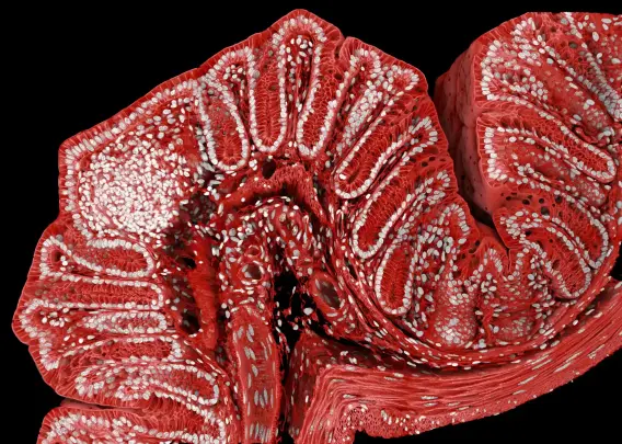
A fluorescently marked mouse colon.
Photograph: Marius Mählen, Koen Oost, Prisca Liberali & Laurent Gelman/Nikon Small World
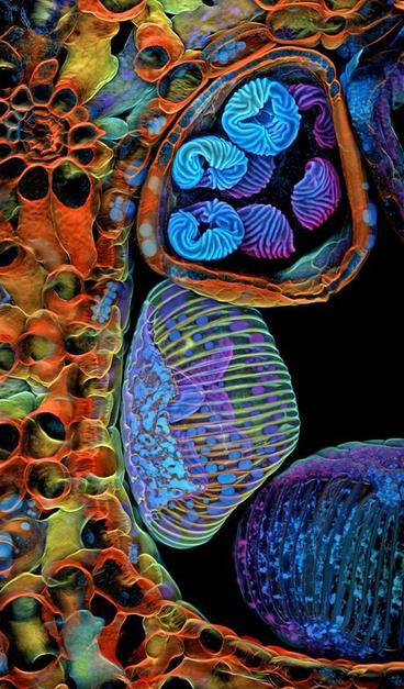
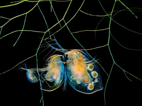
Water fleas (Daphnia) and algae, both vital to underwater ecosystems.
Photograph: Hong Guo/Nikon Small World
Spore sacs (sporangia) of a fern are located on the underside of the fronds, where reproduction happens.
Photograph: Rogelio Moreno Gill/Nikon Small World

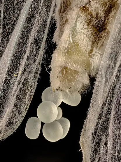
A geometer moth (Geometridae) laying eggs. There are more than 1,270 named Australian species from the Geometridae family, consisting of moths and butterflies.
Photograph: Zhang You/Nikon Small World
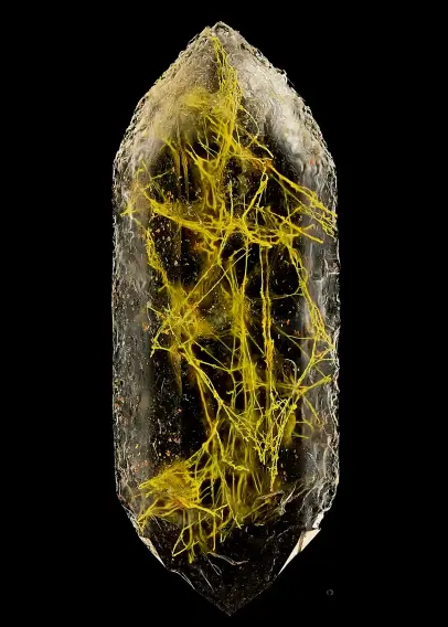
Quartz with biotic goethite filaments.
Photograph: Manfred Heising/Nikon Small World
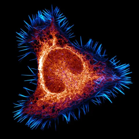
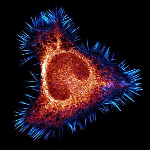
The actin cytoskeleton in cyan, and endoplasmic reticulum in red, show a cancer cell in a mouse’s brain.
Photograph: Halli Lindamood & Eric Vitriol/Nikon Small World