Ricardo Leitão Guerra

@retinography
0 Followers
1 Following
154 Posts
Board certified Ophthalmologist (CBO), fellow of the ICO and BRAVS Member. Sharing reliable scientific information. From Salvador - Brazil
MD, MSc, FICO.

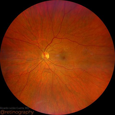
Macular pseudohole

Macular pseudohole is characterized by a steepened foveal contour without a full-thickness defect. On Optical Coherence Tomography (OCT), it shows a narrow, deep foveal pit with intact outer retinal layers, typically caused by a contractile epiretinal membrane (ERM). Differentiating it from a true macular hole is essential, as visual prognosis and management differ significantly. #MacularPseudohole #ERM #OCT #FovealContour #RetinaImaging #retina

https://retinography.org/macular-pseudohole/

Macular pseudohole

Macular pseudohole is characterized by a steepened foveal contour without a full-thickness defect. On Optical Coherence Tomography (OCT), it shows a narrow, deep foveal pit with intact outer retin…

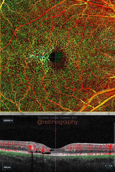
MacTel type 2

In macular telangiectasia type 2 (MacTel), the OCTA depth-enhanced map improves visualization of vascular alterations across different retinal layers. It highlights capillary rarefaction and telangiectatic vessels, particularly in the deep capillary plexus and outer retina. This tool enhances diagnostic accuracy and monitoring of neovascular complications in MacTel. #MacTel #OCTA #DepthEnhancedMap #RetinalVascularChanges #RetinaImaging #retina #oftalmo

https://retinography.org/mactel-type-2-2/

MacTel type 2

In macular telangiectasia type 2 (MacTel), the OCTA depth-enhanced map improves visualization of vascular alterations across different retinal layers. It highlights capillary rarefaction and telan…

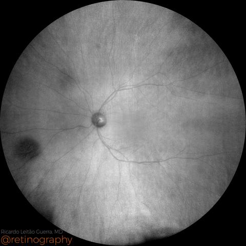
Choroidal nevus

Choroidal nevus is best visualized using near-infrared reflectance (NIR) imaging. Its appearance may vary: it can be hyperreflective in confocal systems or hyporeflective in non-confocal imaging, depending on the imaging modality and melanin density. NIR enhances contrast for detecting and monitoring nevi, especially when they are not clearly visible on color fundus photography or FAF. #ChoroidalNevus #NIR #NearInfraredReflectance #RetinaImaging

https://retinography.org/choroidal-nevus-6/

Choroidal nevus

Choroidal nevus is best visualized using near-infrared reflectance (NIR) imaging. Its appearance may vary: it can be hyperreflective in confocal systems or hyporeflective in non-confocal imaging, …

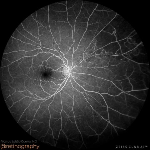
Asymptomatic BRVO

A superior nasal branch retinal vein occlusion (BRVO) is usually asymptomatic, as the macula is typically spared. These cases are often identified incidentally during routine examination. Fluorescein angiography (FA) reveals localized capillary nonperfusion in the affected quadrant, indicating retinal ischemia. Despite the absence of symptoms, close follow-up is essential to monitor for neovascular complications. #BRVO #FluoresceinAngiography

https://retinography.org/asymptomatic-brvo/

Asymptomatic BRVO

A superior nasal branch retinal vein occlusion (BRVO) is usually asymptomatic, as the macula is typically spared. These cases are often identified incidentally during routine examination. Fluoresc…

Proliferative diabetic retinopathy

In proliferative diabetic retinopathy (PDR), neovascularization elsewhere (NVE) refers to abnormal blood vessel growth in the midperiphery or periphery of the retina. The green channel enhances contrast, making NVE more visible against the retinal background. Identifying NVE early is crucial for guiding treatment, such as panretinal photocoagulation (PRP) or anti-VEGF therapy, to prevent complications like vitreous hemorrhage. #PDR

https://retinography.org/proliferative-diabetic-retinopathy-4/

Proliferative diabetic retinopathy

In proliferative diabetic retinopathy (PDR), neovascularization elsewhere (NVE) refers to abnormal blood vessel growth in the midperiphery or periphery of the retina. The green channel enhances co…

Proliferative Diabetic Retinopathy

In proliferative diabetic retinopathy (PDR), subhyaloid hemorrhage results from fragile neovascularization bleeding into the preretinal space. The green channel enhances contrast, improving hemorrhage visualization on fundus imaging. Optical Coherence Tomography (OCT) shows a well-demarcated, hyperreflective hemorrhagic pocket between the retina and the posterior hyaloid face, which can affect visual acuity depending on its location.…

https://retinography.org/proliferative-diabetic-retinopathy-3/

Proliferative Diabetic Retinopathy

In proliferative diabetic retinopathy (PDR), subhyaloid hemorrhage results from fragile neovascularization bleeding into the preretinal space. The green channel enhances contrast, improving hemorr…

AMD: Soft Drusen

In age-related macular degeneration (AMD), soft confluent drusen appear as yellowish deposits on true color imaging, often coalescing into larger lesions. Optical Coherence Tomography (OCT) reveals these drusen as elevations of the retinal pigment epithelium (RPE) with underlying hyporeflective spaces. The presence of large, confluent drusen increases the risk of progression to advanced AMD, making multimodal imaging essential for monitoring. #AMD

https://retinography.org/amd-soft-drusen-3/

AMD: Soft Drusen

In age-related macular degeneration (AMD), soft confluent drusen appear as yellowish deposits on true color imaging, often coalescing into larger lesions. Optical Coherence Tomography (OCT) reveal…

AMD: Outer retinal tubulations

In age-related macular degeneration (AMD), outer retinal tubulations (ORT) can present unique patterns on en-face OCT, often resembling intraretinal fluid (IRF), leading to potential misdiagnosis. In the presented case, en-face imaging shows ORT as hyporreflective interconnected branching networks extensions  surrounded by a hyperreflective borders, while dark areas without hyperreflective borders at the periphery represent subretinal…

https://retinography.org/amd-outer-retinal-tubulations/

AMD: Outer retinal tubulations

In age-related macular degeneration (AMD), outer retinal tubulations (ORT) can present unique patterns on en-face OCT, often resembling intraretinal fluid (IRF), leading to potential misdiagnosis.…

Central Serous Chorioretinopathy

In Central Serous Chorioretinopathy (CSC), serous retinal detachment and serous pigment epithelial detachment (PED) are hallmark findings. Optical Coherence Tomography (OCT) reveals a hyporeflective space beneath the neurosensory retina and PEDs, often associated with focal RPE abnormalities. The leakage site at the RPE can be identified on OCT as a point of discontinuity or hypertransmission, guiding diagnosis and management. #CSC

https://retinography.org/central-serous-chorioretinopathy-10/

Central Serous Chorioretinopathy

In Central Serous Chorioretinopathy (CSC), serous retinal detachment and serous pigment epithelial detachment (PED) are hallmark findings. Optical Coherence Tomography (OCT) reveals a hyporeflecti…

Untitled

In degenerative myopia, situs inversus refers to the tilted insertion of the optic disc, commonly associated with retinal and choroidal changes. Optical Coherence Tomography (OCT) B-scan often shows tilted scans due to the oblique orientation of posterior pole structures. This tilting can distort retinal layer visualization, requiring careful interpretation to assess associated pathologies like staphyloma, choroidal thinning, or macular neovascularization.…

https://retinography.org/2226-2/

(no title)

In degenerative myopia, situs inversus refers to the tilted insertion of the optic disc, commonly associated with retinal and choroidal changes. Optical Coherence Tomography (OCT) B-scan often sho…