Xiaoxuan Jia et al. investigated the correlation of the mitotic index of gastric gastrointestinal stromal tumors with CT-identified morphological and first-order #radiomic features, finding that the invasive margin could be the sole independent CT high-risk morphological feature for 1–5 cm gGISTs after tumor size-based subgroup analysis.

CT assessed morphological features can predict higher mitotic index in gastric gastrointestinal stromal tumors - European Radiology
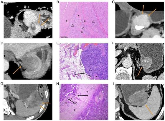
Objectives To investigate the correlation of the mitotic index (MI) of 1–5 cm gastric gastrointestinal stromal tumors (gGISTs) with CT-identified morphological and first-order radiomics features, incorporating subgroup analysis based on tumor size. Methods We enrolled 344 patients across four institutions, each pathologically diagnosed with 1–5 cm gGISTs and undergoing preoperative contrast-enhanced CT scans. Univariate and multivariate analyses were performed to investigate the independent CT morphological high-risk features of MI. Lesions were categorized into four subgroups based on their pathological LD: 1–2 cm (n = 69), 2–3 cm (n = 96), 3–4 cm (n = 107), and 4–5 cm (n = 72). CT morphological high-risk features of MI were evaluated in each subgroup. In addition, first-order radiomics features were extracted on CT images of the venous phase, and the association between these features and MI was investigated. Results Tumor size (p = 0.04, odds ratio, 1.41; 95% confidence interval: 1.01–1.96) and invasive margin (p < 0.01, odds ratio, 4.55; 95% confidence interval: 1.77–11.73) emerged as independent high-risk features for MI > 5 of 1–5 cm gGISTs from multivariate analysis. In the subgroup analysis, the invasive margin was correlated with MI > 5 in 3–4 cm and 4–5 cm gGISTs (p = 0.02, p = 0.03), and potentially correlated with MI > 5 in 2–3 cm gGISTs (p = 0.07). The energy was the sole first-order radiomics feature significantly correlated with gGISTs of MI > 5, displaying a strong correlation with CT-detected tumor size (Pearson’s ρ = 0.85, p < 0.01). Conclusions The invasive margin stands out as the sole independent CT morphological high-risk feature for 1–5 cm gGISTs after tumor size-based subgroup analysis, overshadowing intratumoral morphological characteristics and first-order radiomics features. Key Points Question How can accurate preoperative risk stratification of gGISTs be achieved to support treatment decision-making? Findings Invasive margins may serve as a reliable marker for risk prediction in gGISTs up to 5 cm, rather than surface ulceration, irregular shape, necrosis, or heterogeneous enhancement. Clinical relevance For gGISTs measuring up to 5 cm, preoperative prediction of the metastatic risk could help select patients who could be treated by endoscopic resection, thereby avoiding overtreatment.