I've not been here much lately, but here's a bit more about our paper just out in the Journal of Cell Biology, “Presynapses contain distinct actin nanostructures” (Twitter trained me to come up with short titles 🤏 and long threads 🧵, which are also doable in Mastodon I hope)
The article is available here:
https://rupress.org/jcb/article-abstract/222/10/e202208110/276185/Presynapses-contain-distinct-actin1/23
Presynapses contain distinct actin nanostructures | Journal of Cell Biology | Rockefeller University Press
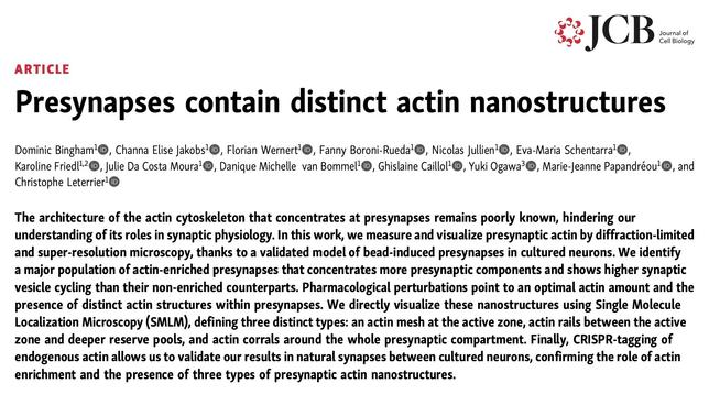
Bingham et al. use bead-induced isolated presynapses and CRISPR-mediated tagging of endogenous actin in neurons coupled to super-resolution microscopy to reveal
I want to highlight the work that went into the revision of the manuscript, crediting the contribution of lab members and collaborators, as well as give an overview of the Review Commons process we used (see
https://www.reviewcommons.org/) 2/23

Homepage
Improve your paper and streamline publication through journal-independent peer-review.
We previously shared this work as a bioRxiv preprint in May last year, as the main work of Dominic Bingham who graduated in July 2022 (see Twitter thread:
https://twitter.com/christlet/status/1527183879643201536?s=20) 3/23

Christophe Leterrier on Twitter
“🚌 Next stop on the NeuroCyto lab axonal journey: the presynapse🚏 Ever wondered how is presynaptic actin organized beyond the blobs seen by diffraction-limited microscopy? We certainly did! So why not do some STORM on it? https://t.co/0HFfmgQcm0 (1/6)”
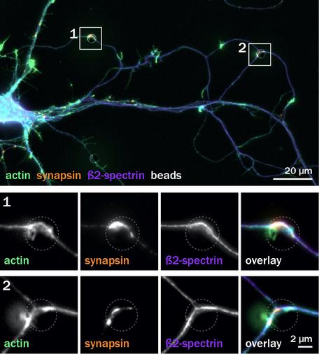
In this work, we used beads to induce isolated presynapses along axons of cultured neurons, making it possible to directly visualize and quantify presynaptic actin without interference from the concentrated postsynaptic actin 4/23
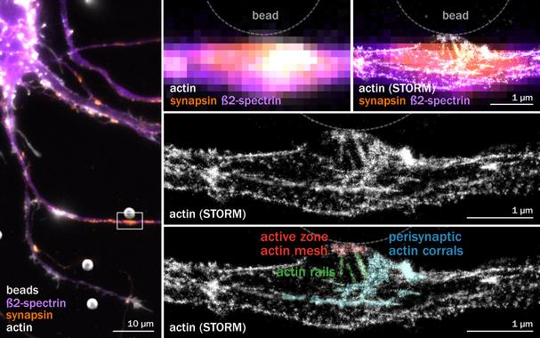
We directly visualized the selective enrichment of actin at presynapses, and could get good SMLM images of presynaptic actin at the nanoscale within bead-induced presynapses 5/23
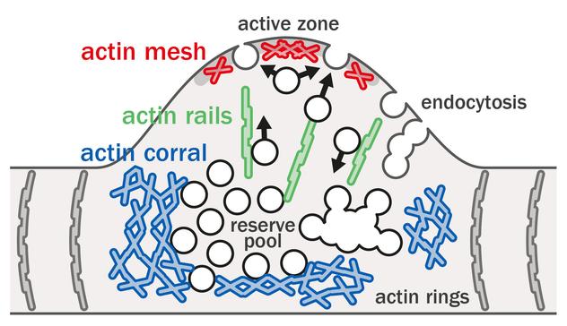
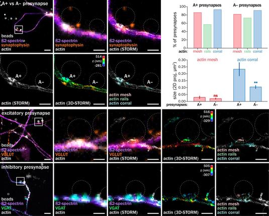
This led us to define three types of presynaptic actin nanostructures: a faint actin mesh at the active zone, actin rails within the presynapse, and dense perisynaptic actin corrals 6/23
I was curious to try Review Commons and have our work openly reviewed upstream of journal submission so we submitted our preprint there, and the reviews were posted next to the preprint in July 2022 7/23
The reviewers found the work interesting and important, but they wanted 1) more controls about the bead-induced presynapses, 2) better characterization of the actin structures at the nanoscale, and 3) confirmation in “real” synapses between two neurons 8/23
We had 4 weeks to draft a detailed revision plan, then submitted the preprint together with reviews and revision plan to journals of the Review Commons consortium, with assurance they would reply within 10 days that they would consider a revised manuscript for publication 9/23
I was over the moon when J Cell Biol editor Andrea Marat gave us a positive answer. So off we go in September to revise the manuscript and address the three points listed above - it was a lot of work! 10/23
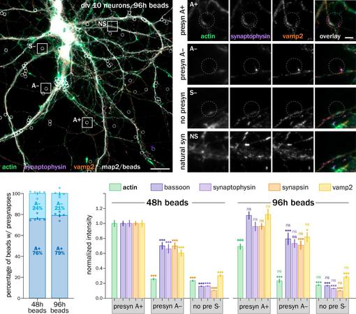
Marie-Jeanne Papandréou took care of all the bead-induced presynapse quantitative analyses with Fanny Boroni-Rueda, measuring hundreds more synapses to confirm that the bead-induced presynapses were indeed mature, and very similar to natural synapses 11/23
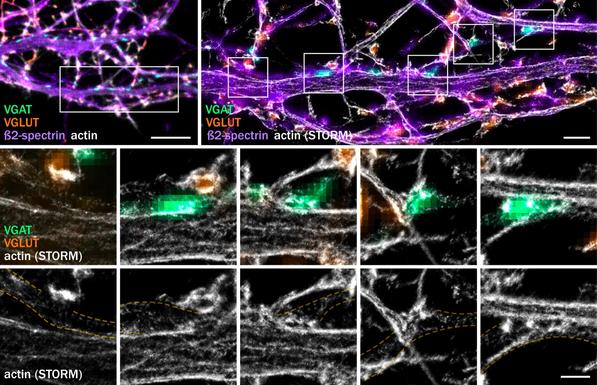
On the SMLM front, we were lucky to have two great Master students joining us. Channa Jakobs from the Netherlands worked on STORM of actin (with DECODE processing), detailing how presynaptic actin is hidden by postsynaptic actin in excitatory and inhibitory natural synapses 12/23
Channa also showed that the actin mesh, rails and corrals are present in all bead-induced presynapses, with variations in the size of the perisynaptic corrals. Mesh, rails and corrals are also found in both excitatory and inhibitory induced presynapses 13/23
Meanwhile, Master student Eva Schentarra from Germany skillfully worked with Karoline Friedl on our Abbelight setup to obtain 2-color spectral STORM images of actin and Arp2/3, showing that the actin nucleator preferentially localizes to the mesh and corrals, but not rails 14/23
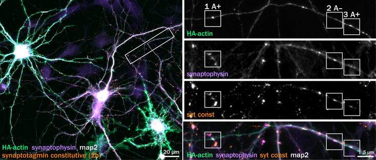
Finally, visualizing these nanostructures in the presynaptic side of a natural synapse was a large project in itself. Simple transfection of actin probes like LifeAct cannot provide good-enough SMLM images, so we developed CRIPSR-based tagging of endogenous actin in neurons 15/23
This was done by Nicolas Jullien in collaboration with Yuki Ogawa from Matt Rasband's lab, who developed an AAV-based variant of HiUGE for rat hippocampal neurons 16/23
With these tools, Dominic Bingham last experiments in the lab showed that natural presynapses expressing HA-tagged actin had a selective enrichment in actin similar to bead-induced ones, which was also linked to a higher level of presynaptic components and vesicular cycling 17/23
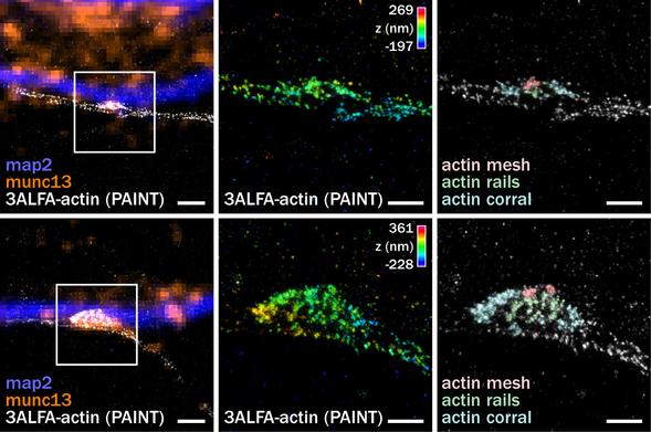
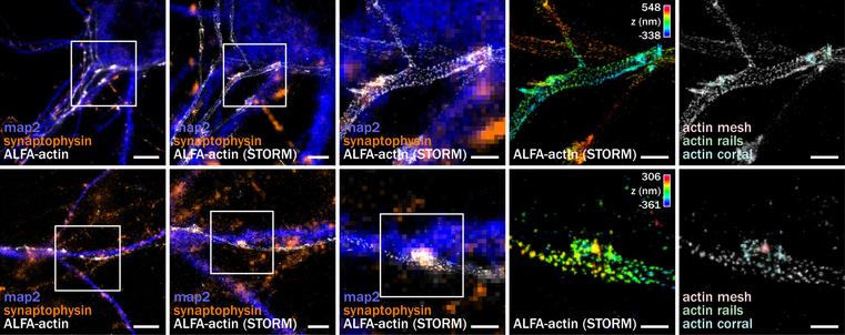
The key experiment was then to image these CRISPR-tagged natural synapses to confirm the presence of the presynaptic actin nanostructures. This required a lot of work for Nicolas to design various tags and for Channa to perform SMLM on tagged neurons 18/23
Difficulty here was to get enough localizations to delineate the structures, but with a good enough precision to not blur things too much: small probe, with a lot of blinking events. We tested different tags labeled with antibodies or nanobodies, imaged by PAINT and STORM 19/23
In the end the best results were obtained using an ALFA tag labeled with primary and secondary antibodies, or a triple ALFA tag with an anti-ALFA PAINT nanobody. In both cases, we could visualize the actin mesh, rails and corrals in tagged presynapses! 20/23
So about a year after posting the preprint, we were ready to resubmit to
@JCellBiol. Reviewers were happy, and things went swiftly after that. This journal is very special to me because of all the landmark neuronal cell biology papers it has published over the decades 21/23
I liked the open
@ReviewCommons process, and if you are interested, you can check the reviews, detailed revision plan and rebuttal which are available next to the article 22/23
Finally, thanks to everyone who worked for months/years on this project, and to the funders that allowed to develop an ambitious new direction for the lab. A special thanks to Subhojit Roy for hooking me up with the bead-induced presynapses 🙌
https://rupress.org/jcb/article-abstract/222/10/e202208110/276185/Presynapses-contain-distinct-actin23/23 - Thanks for reading the whole thread!
Presynapses contain distinct actin nanostructures | Journal of Cell Biology | Rockefeller University Press
Bingham et al. use bead-induced isolated presynapses and CRISPR-mediated tagging of endogenous actin in neurons coupled to super-resolution microscopy to reveal
@christlet beautiful
#microscopy as always. Great to see you around here.