πŸ§ πŸ’πŸπŸ¦˜πŸ¦™πŸ¦ŒπŸ·πŸ»β€β„οΈπŸ¦«πŸ¦πŸ‘πŸ‡πŸˆπŸ¦”πŸ•πŸ¦‡πŸ¦­πŸ¦₯πŸ¦“
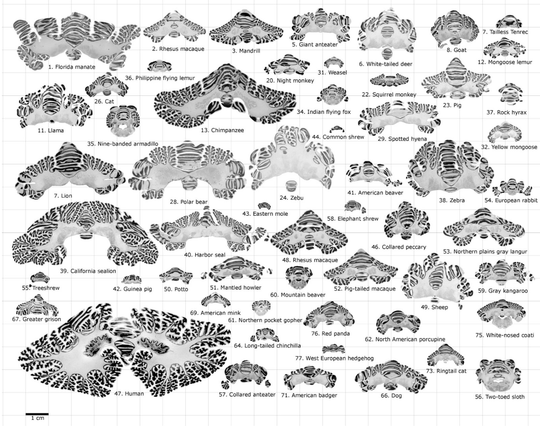
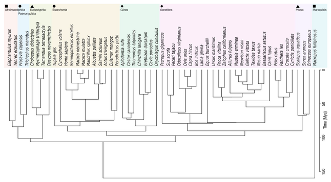
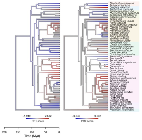
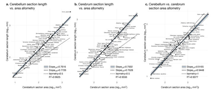
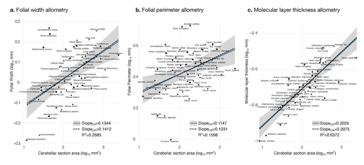
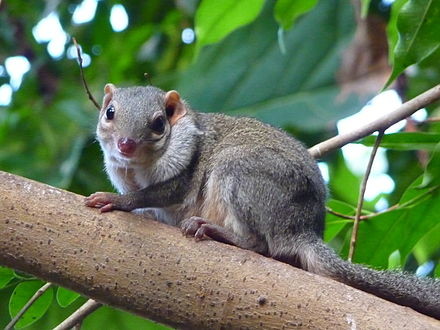
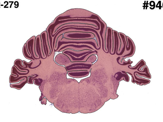
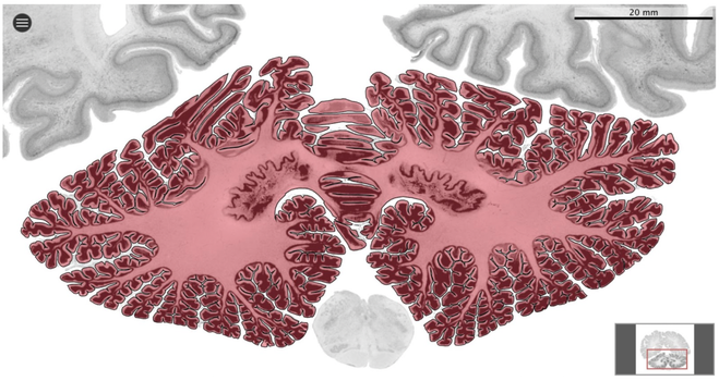
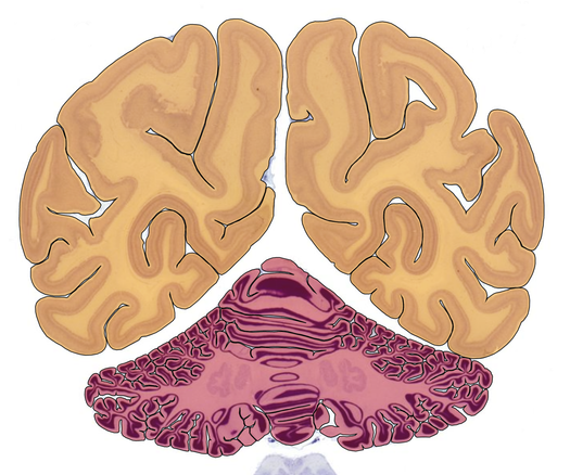
We’ve been diving into the mesmerising anatomical diversity and evolution of cerebellar folding across 56 mammalian species with @r3rt0 Nicolas Traut @AleAliSousa @sofievalk
https://www.biorxiv.org/content/10.1101/2022.12.30.522292v1

Check it out in a short tooting thread πŸ”½

@r3rt0 @AleAliSousa @sofievalk

2
We used our Web tool MicroDraw to visualise and segment the histological data online, and created tools to study the geometry of cerebellar folia and to estimate the thickness of the molecular layer.

@r3rt0 @AleAliSousa @sofievalk

3
Phylogenetic comparative methods revealed that the evolution of cerebellar and cerebral neuroanatomy follows a stabilising selection process.

@r3rt0 @AleAliSousa @sofievalk

4
Cerebellar size and folding correlate strongly with cerebral size and folding.
Ancestral character state estimations showed that size and folding of the cerebrum and cerebellum increase and decrease concertedly through evolution.

@r3rt0 @AleAliSousa @sofievalk

5
The cerebellar and cerebral cortices are disproportionately larger than their volumes, as shown by their hyper-allometry. The cerebellum is slightly but statistically significantly hypo-allometric compared to the cerebrum.

@r3rt0 @AleAliSousa @sofievalk

6
Folial width, folial perimeter and the thickness of the molecular layer increase slightly with cerebellar size, largely conserved when compared with changes in total cerebellar size, as revealed by the small allometric slopes.

@r3rt0 @AleAliSousa @sofievalk

7
Our results confirm the strong correlation between cerebral and cerebellar volumes across species, and we extended these results to show that the same strong relationship holds for cerebellar folding: larger cerebella appear to be disproportionately more folded than smaller ones.

@r3rt0 @AleAliSousa @sofievalk

8
Comparing the allometric relationships between section length and area suggests that a similar process lies behind the folding of the cerebrum and the cerebellum, that can be explained by buckling and where wavelength of folding depends on cortical thickness.

@r3rt0 @AleAliSousa @sofievalk

9
We work collaboratively online! Please reach out and come join us at any time! ❀️ πŸ’ πŸ’ πŸ’

@k4tj4 @r3rt0 @AleAliSousa @sofievalk
Very cool work and cool data!!!! πŸ‘πŸ‘ are you guys planning to look at the cytoarchitecture on your histological data ? 🀩

@kepkeeloh @r3rt0 @AleAliSousa @sofievalk

Yes! Next up! The detection of the molecular layer in the histological data was a 1st step (allowed us to make sure that the vectorial and pixel data are well aligned, and that we can combine both). Roberto is much more advanced in the analysis of his ferret data, doing 3d profiles and histology-based surface reconstructions, etc. Wanna join our adventure 🀩 ? It works across the ocean πŸ’ž First step will be more segmenting... πŸ˜…

@k4tj4 @r3rt0 @AleAliSousa @sofievalk

Awesome!!!! I’m curious to look at the nissl sections of the various mammal species and see how the various brain areas compare with les primates 🀩!!

By segmentation you mean the identifying of the various layers within the cortex right? We can see the individual neurons on brain box?

Great start to 2023!!!!

@k4tj4 @r3rt0 @AleAliSousa @sofievalk what about the deep cerebellar nuclei?

@k4tj4 @r3rt0 @AleAliSousa @sofievalk

Such a very beautiful work, congrats for it! I have recently watched a lecture of a Brazilian professor (link to his contact below) that works in a similar research line. He uses math modelling to understand the dynamics of brain folding. Check it out:

https://www.if.ufrj.br/docentes/bruno-motta/

@inaciomdrs @k4tj4 @AleAliSousa @sofievalk Yes! We know Bruno's work well. He's doing some amazing stuff.
@k4tj4 @r3rt0 @AleAliSousa @sofievalk A beautiful study. Were you expecting this correlation from a computational perspective? I have always assumed this would be the case (since cortex and cbm carry out something like argmax(Wr) where W is stored in the cbm and r is produced by the cortex http://wiki.glennersterlab.com/index.php?title=The_big_idea). It is great to see this mapped out across species.
The big idea - A conversation about the brain

@ag3dvr @k4tj4 @AleAliSousa @sofievalk
Thank you Andrew! And thank you for the link to the big question!
We were expecting a correlation, but didn't have a hypothesis for the scaling coefficient. It could be that you need proportionally more cerebellum with progressively larger cerebrums (scaling>1), or that they need to stay fully proportional (scaling~1). What we observed was a scaling slightly, but statistically significantly smaller than 1. What would have been your guess?

@r3rt0 @k4tj4 @AleAliSousa @sofievalk
Great to be in a conversation about this – my first on Mastodon!

I am afraid I don’t think I can contribute to the subtlety of the question you are asking (cbm:cx ratio of slightly greater, slightly less or equal to 1:1). But I am pleased to hear that it is close to 1:1 throughout evolution. I will try and say a bit more about why that seems to make sense from a computational perspective. 1/n

@r3rt0 @k4tj4 @AleAliSousa @sofievalk

Here is a video of me trying to explain why neural nets and the cerebellum are similar:
https://www.youtube.com/watch?v=NiRPq11wA-A
Two changes in evolution:
(i) dimensionality of the input (by assumption, cortical) vector and of the stored (by assumption, cerebellar) vectors
(ii) the length of paths through that space to achieve rewards.
I make some mistakes in that video, eg a policy network is _all_ the state-contingent actions (Ο€(a|x)), not just a single instance. 2/n

BMVA Symposium-VIIHM-Andrew Glennerster β€œPolicy networks with and without brains”

YouTube

@r3rt0 @k4tj4 @AleAliSousa @sofievalk

Here is the paper where I get that right, but I do not talk about evolution:
https://royalsocietypublishing.org/doi/10.1098/rstb.2021.0448 3/n

Understanding 3D vision as a policy network | Philosophical Transactions of the Royal Society B: Biological Sciences

It is often assumed that the brain builds 3D coordinate frames, in retinal coordinates (with binocular disparity giving the third dimension), head-centred, body-centred and world-centred coordinates. This paper questions that assumption and begins to ...

Philosophical Transactions of the Royal Society B: Biological Sciences

@r3rt0 @k4tj4 @AleAliSousa @sofievalk

In terms of argmax(Wr), cortical size relates to n (the length of r and the first dimension (width) of W). Cerebellar size relates to m (the second dimension (height) of W). n and m are, in theory, free to vary independently but clearly in practice they will co-vary. Higher dimensional spaces allow longer paths to be traversed through that space to achieve rewards, hence m grows with n (more Purkinje cells with more cortex). 4/n

@r3rt0 @k4tj4 @AleAliSousa @sofievalk

But, as I say, I don’t see that that helps with your question of whether you should expect slightly greater, slightly less or equal to 1:1. I would hazard a guess that you don't need quite the proportional increase in cbm (m) as cortex (n) increases as you will be able to programme a rich behavioural repertoire in the available high dimensional space... but others would be better placed to comment. 5/n

@r3rt0 @k4tj4 @AleAliSousa @sofievalk

Fig 1 from the Phil Trans paper cited above, drawing the link between deep neural nets and the cerebellum. 6/n

@r3rt0 @k4tj4 @AleAliSousa @sofievalk

... but also why deep neural nets 'fall flat on their face'.... but that is another topic.
https://www.youtube.com/watch?v=oDLtPY1e9bk. 7/7

TVG draft talk 28jul2022

YouTube

@r3rt0 @k4tj4 @AleAliSousa @sofievalk

PS. Talking with colleagues who study ants, their best guess of the equivalent of the cerebellum was the mushroom bodies. Every nervous system has to have some equivalent (a W and an r). Is that your best guess too? 8/8