I've got a protein gel mystery.
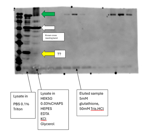
I took exactly the same frozen cell lysate and resuspended it in two different buffers and the western blot looks completely different.
In PBS +triton, I have a crisp band of the protein of interest at the top (green), a known cross-reacting band, and a small band (yellow ??), in HEK5G+chaps the band of interest is blurry and the small band is missing.
What is happening here?