For
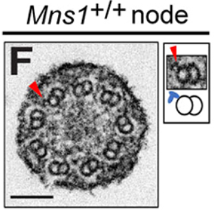
#EMmonday, we’re highlighting an EM favourite, cilia! Hao Lu, Wang Kyaw Twan, Yayoi Ikawa, Vani Khare, Hiroshi Hamada, Sudipto Roy & colleagues find that ODAs are lost from Mns1 mutant mouse (9+0) nodal cilia.
📖
@Dev_journal:
https://journals.biologists.com/dev/article/151/14/dev202737/361132/Localisation-and-function-of-key-axonemalIt’s
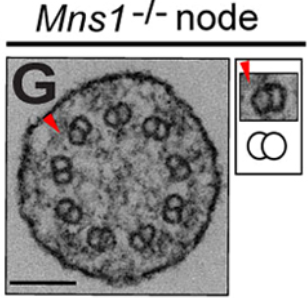
#EMmonday and we’re highlighting
#zebrafish photoreceptors! Hung-Ju Chiang, Ichiro Masai and colleagues find that the axoneme is absent in payday mutant photoreceptors.
📖
@DMM_Journal:
https://journals.biologists.com/dmm/article/17/7/dmm050618/361166/Male-germ-cell-associated-kinase-is-required-forOn our return to
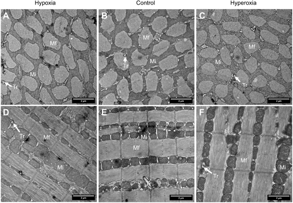
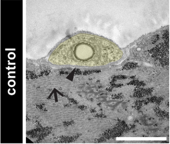
#EmMonday, we’re highlighting electron micrographs of Drosophila larval NMJ from Briana Christophers, et al. The images, taken with the assistance of the Weil Cornell Medicine Microscopy Core Facility, show defects in post-synaptic actin organisation and the synaptic cleft.
📖
@Dev_journal:
https://journals.biologists.com/dev/article/151/13/dev202558/359693/Muscle-cofilin-alters-neuromuscular-junctionHappy
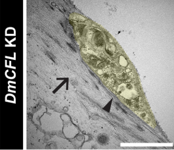
#EMmonday! This week we’re zooming in on Arabidopsis embryos using SEM. Yosapol Harnvanichvech, Cecilia Borassi, Joris Sprakel and Dolf Weijers and colleagues identify a proteinaceous envelope that encapsulates the early Arabidopsis embryo.
@Dev_journal:
https://journals.biologists.com/dev/article/150/22/dev201943/334678/An-elastic-proteinaceous-envelope-encapsulates-the
An elastic proteinaceous envelope encapsulates the early Arabidopsis embryo
Summary: The early Arabidopsis embryo is surrounded by a proteinaceous envelope that is distinct from the cuticle and embryo sheath.
It’s
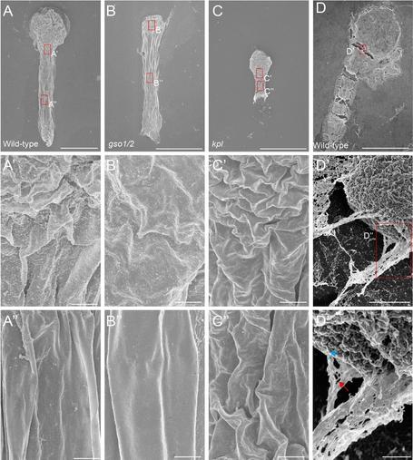
#EMmonday and we’re highlighting Drosophila eyes imaged using SEM from Akanksha Onkar, Subramaniam Ganesh and colleagues. They find that increased brain glycogen ameliorates Huntingdon’s disease phenotypes.
📖
@DMM_Journal:
https://journals.biologists.com/dmm/article/16/10/dmm050238/333679/Increase-in-brain-glycogen-levels-ameliorates
Increase in brain glycogen levels ameliorates Huntington's disease phenotype and rescues neurodegeneration in Drosophila
Summary: Whereas healthy brain contains very little glycogen, aged brains and brains of people with neurodegenerative disorders tend to accumulate glycogen. We demonstrate here that, in the Drosophila model of Huntington's disease, enhancement of glycogen synthesis in fly brain ameliorated the disease phenotype through activation of auto-lysosomal functions.
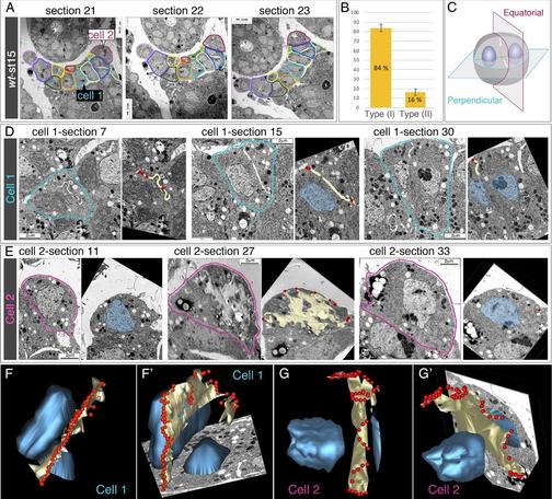
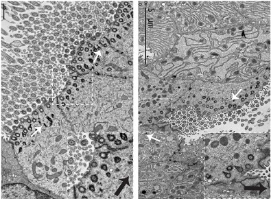
On
#EMmonday we’re highlighting slit assembly in Drosophila nephrocytes. Marta Carrasco-Rando, Mar Ruiz-Gómez and colleagues @CBMSO_CSIC_UAM discover that an acytokinetic cell division breaks membrane symmetry during slit diaphragm assembly.
📖
@Dev_journal:
https://journals.biologists.com/dev/article/150/18/dev201708/328448/An-acytokinetic-cell-division-creates-PIP2#OpenAccess
An acytokinetic cell division creates PIP2-enriched membrane asymmetries leading to slit diaphragm assembly in Drosophila nephrocytes
Summary: An acytokinetic cell division breaks the initial membrane symmetry in embryonic garland nephrocytes creating PIP2-enriched membrane microdomains suitable for slit diaphragm assembly.
Happy #EMmonday! This week we are highlighting research from Riley Cooney, Eszter Vladar and colleagues. They find that DKK3 and WNT4 work together to enable a switch from canonical to non-canonical Wnt signalling during multiciliogenesis.
https://journals.biologists.com/jcs/article/136/16/jcs260807/326574/A-WNT4-and-DKK3-driven-canonical-to-noncanonical

A WNT4- and DKK3-driven canonical to noncanonical Wnt signaling switch controls multiciliogenesis
Summary: Identification of novel regulators of the Wnt pathway that control multiciliated cell formation in the airway epithelium.
This
#EMmonday we are looking at axoneme organisation with Tomohiro Kubo, Toshiyuki Oda and colleagues. They find that that the glutamate-rich C-terminal tails of α- and β-tubulin play essential, and distinct, roles in ciliary motility and assembly.
Highlight:
https://journals.biologists.com/jcs/article/136/16/e136_e1601/325932/Roles-of-ciliary-tubulin-PTMs-in-Chlamydomonas

Roles of ciliary tubulin PTMs in Chlamydomonas reinhardtii
The cytoskeletal core of cilia and flagella, called the axoneme, is composed of microtubules (MTs), the dynamics of which can be dramatically influenced by post-translational modifications (PTMs) to tubulin C-terminal tails. In mammals, the presence of many tubulin genes, protein isoforms and potential combinations of PTMs make understanding the roles each might have in ciliary function extremely challenging. The biflagellate unicellular algae Chlamydomonas reinhardtii, however, possesses only four tubulin genes, which encode two pairs of identical α- and β-tubulins. In this study (Kubo et al., 2023), Toshiyuki Oda and colleagues take advantage of the simplicity of Chlamydomonas tubulin expression to explore the ciliary roles of tubulin PTMs by mutating key residues to block either glutamylation or glycylation on α- or β-tubulin tails, respectively. Loss of glutamylation of α-tubulin causes severe loss of cell motility despite having no clear effect on axoneme structure, suggesting that it influences the physical properties of ciliary MTs. In contrast, blocking glycylation of β-tubulin produces truncated, nonmotile flagella lacking central axonemal MTs, resembling other Chlamydomonas mutants for the MT-severing ATPase katanin. These findings thus illuminate distinct but crucial functions for both PTMs in ciliary motility, which might also contribute to understanding pathogenic mechanisms in human ciliopathies.
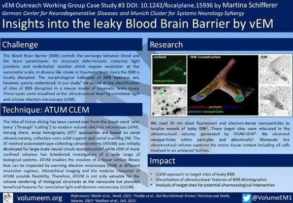
We’re doubling the fun this #EMmonday as we also get to highlight the latest #vEM case study on FocalPlane from Martina Schifferer and colleagues! They used ATUM CLEM to visualise the ultrastructural features of BBB disintegration.
vEM Outreach WG Case Study #3
https://focalplane.biologists.com/2023/08/04/insights-into-the-leaky-blood-brain-barrier-by-vem/

Insights into the leaky Blood Brain Barrier by vEM - FocalPlane
Insights into the leaky Blood Brain Barrier by vEM - Blog series